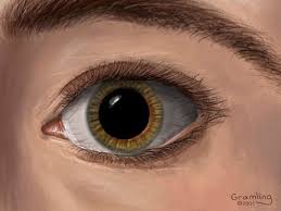
It is a bone structure applied to the front and bottom of the head . Its upper and lower limits are the forehead and chin , and the lateral limits are the ears . The soft tissues of the face express the emotions of the individual, by the action of the muscles of mimicry . Likewise, facial appearance is an essential element of the identity of each human being.
Summary
[ hide ]
- 1 Structure
- 2 Bones of the face
- 3 Muscles of the face
- 1 Masseter muscle
- 2 External or lateral pterygoid muscle
- 3 Internal or medial pterygoid muscle
- 4 Cutaneous muscles of the face
- 5 Muscles of the eyelids and eyebrows
- 5.1 Occipitofrontal muscle
- 5.2 Orbicularis oculi muscle
- 5.3 Superciliary muscle
- 6 Nose muscles
- 6.1 Pyramidal muscle of the nose or procedure
- 6.2 Transverse muscle of the nose
- 6.3 Myrtiform muscle of the nose
- 6.4 Dilator muscle of the nostrils of the nose
- 7 Muscles of the lips
- 7.1 Deep dilator muscles
- 7.2 Superficial dilator muscles
- 7.3 Constrictor muscles
- 4 Sources
Structure
The face is made up of:
- Eyebrows
- Eyes
- Nose
- Cheek
- Mouth
- Lips
- Teeth
- Skin
- Chin
Over thirty pairs of muscles are implanted on the hard surface of the bones , of various shapes and functions, thick and strong, like the masseter , others are thin and small, like the one that raises the corner of the lips and the one that dilates the wing. of the nose , among others.
Of the five sense organs , four are in the head , and more specifically in the face; the eye , the ear , the smell and taste. Although the fifth sense, touch , is also widely represented, with a very sensitive area on the lips .
The facial mass is organized in two jaws , one upper and one lower. The upper jaw is made up of thirteen bones , all of which are pairs except one, the vomer . Instead, a single bone constitutes the lower jaw: the lower jaw.
Schematically:
- Upper jaw.
- Upper jaws.
- Own bones of the nose.
- Palatine.
- Lower turbinates.
- The vomer.
- Lower jaw.
- Lower jaw.
- Chewing muscles.
- Muscles of the eyelidsand eyebrows .
- Nose muscles.
- Pavilion muscles.
- Lip muscles.
Bones of the face
See Bones of the face
Muscles of the face
The muscles of the head are divided into chewing muscles and skin muscles. The chewing muscles are divided into 4 pairs and are: the temporal, the masseter, the external pterygoid and the internal pterygoid.
Masseter muscle
- Concept: The term is derived from the Greek masseter, chew and defines its main function. The masseter muscle largely covers the branch of the lower jaw and in turn is covered by maseteric or parotideomaseteric fascia.
- Location: It extends from the zygomatic arch to the external face of the ascending branch of the lower jaw.
- Origin: It originates from the tendon fascicles of the anterior and middle portions of the zygomatic arch. It originates in the middle and posterior segments of the zygomatic arch and almost always runs vertically downwards, to the external face of the branch of the mandible.
- Insertions: The muscle is inserted above in the zygomatic arch and upper jaw, and below almost all the lateral or external aspect of the branch of the mandibleand in a lower part of the coronoid process.
- Innervation: It comes from the masseterine branch of the N. inferior maxillary division of the N. trigeminal.
- Irrigation: It comes from the maseteric and transverse arteriesof the face.
- Action: The contraction of the masseter muscle produces the constriction of the jaws. Lift your jaw; the superficial portion participates in the protrusion of the mandible forward. Its most relevant contribution is manifested in the act of chewing.
External or lateral pterygoid muscle
- Concept: The term originates from the Greek ptergion, fin and describes the shape of the muscle.
- Location: It is located in the infratemporal fossa below the temporal muscleand above the pterygoid lamina and the internal or medial pterygoid muscle.
- Origin: It originates from two beams, upper or sphenoid beam, the same is born:
- From the horizontal part of the external face of the greater wing of the sphenoid.
- From the spheno-temporal ridge.
- From the upper third or fourth of the external aspect of the external wing of the pterygoid process.
- Lower beam or pterygoid, which is born:
- In the three-fourths or the lower two-thirds of the external aspect of the external wing of the pterygoid process.
- On the external face of the palatal pyramidal process.
- In the adjacent part of the tuberosity of the upper jaw.
- Insertion: The two fleshy bodies are immediately confused and end at the same time by fleshy fibers and small tendon fascicles:
- At the anterior border of the interarticular fibrocartilage.
- In the antero-internal fossa of the neck of the maxillary condyle.
- Innervation: It comes from the lateral pterygoid branch of the division of n. lower jaw of n. trigeminal the n. buccal emerges between the two bundles or bellies of this muscle.
- Irrigation: It comes from a. facial.
- Action: Bring the jaw forward in the protrusion movement and direct the chin to the opposite side. When the muscles contract simultaneously, the jawis brought forward. The muscle also participates in the opening of the mouth , pulling the capsule and disc of the temporomandibular joint and the process that holds the condyle forward.
Internal or medial pterygoid muscle
- Concept: The term originates from the Greek ptergion, fin and describes the shape of the muscle.
- Location: It is located deep in the infratemporal fossa. It partly covers the lower fibers of the external pterygoid muscle and is also partially covered by them.
- Origin: It originates from the tuberosity of the upper jaw and passes through the lower fibers of the external pterygoid muscle at its point of origin. The greater portion arises from the internal face of the pterygoid process and the pyramidal process of the palatine bone. On the entire surface of the pterygoid fossa, except in the posterior part of the internal wall of this fossa and in the scaphoid fossa, parts that are in relation to the external peristaphilin muscles.
- Insertion: The maxillary insertions of this muscle are made by fleshy fibers and by tendon sheets on the inner side of the lower jaw below and behind the mylohyoid canal.
- Innervation: It is crossed to the superficial plane by the inferior lingual and dental nerves, which emerge below the external pterygoid muscle.
- Irrigation: It comes from the alveolar, buccal and facial arteries.
- Action: It is a jaw elevator. The alternate contraction of the two internal pterygoid muscles concur with the teaching movements.
Cutaneous muscles of the face
The skin muscles are those that always have inserts in the skin, and are innervated by the facial nerve. They are characterized by being flat and thin and are located, for the most part around the eye holes, nose and mouth, they have been defined as muscles of expression and for this reason the human being is able to express the different states of mind .
Eyelid and eyebrow muscles
They are four:
- Orbicularis of the eyelids.
Occipitofrontal muscle
- Concept: It is a quadrilateral, digastric muscle, formed by the occipital muscles backwards and the frontal muscles forward, separated by the epicranial fascia.
- Frontal muscles origin: Located in the anterior part of the head and extending from the epicranial fascia.
- Insertion: it is inserted from above, at the anterior edge of the epicranial fascia at its convex edge and ends in the superciliary and interciliary regions.
- Occipital muscles origin: At the posterior edge of the epicranial fascia.
- Insertion: On the upper lip of the upper curved occipital line.
- Innervation: Temporofacial branch of the facial for the frontal muscles and for the occipitals by the occipital nerve.
- Irrigation: Through the frontal, occipital and superficial temporal arteries.
- Action: Tensor epicranial fascia especially the occipitals. The frontal muscles determine the transverse wrinkles on the forehead.
Orbicularis oculi muscle
- Concept: Muscle, which surrounds the palpebral orifice as a ring.
- Origin: Its ciliary portion originates from the orbital aspect of the tear bone. The orbital portion extends around an ellipse and is not interrupted at the palpebral commissure. The palpebral portion originates from the internal palpebral ligament.
- Insertion: At the corners of the eye. In the internal angle, by means of a tendon formed of two parts, one of them a direct tendon, it is inserted in the anterior area of the tarsal lamina and the other, the reflex tendon, is inserted into the lateral palpebral raphe. In the external angle, the muscle fascicles intersect and end in the skin of the frontal region.
- Innervation: Zygomatic branch of the facial.
- Irrigation: Ophthalmic artery.
- Action: The ciliary or lacrimal portion influences lacrimal secretion, sucking and compressing. The orbital portion closes the eyelids with energy as when trying to protect the eye from the entry of foreign elements. The eyelid portion gently closes the eyelids and regulates the finer movement of the eyelids.
Superciliary muscle
- Concept: Musclesuperciliar or corrugator or brow depressor muscle.
- Origin: It is a short muscle extended over the internal part of the superciliary arch.
- Insertion: It is born in the internal part of the superciliary arch; it is directed upwards and outwards and at the level of the supraorbital hole the skin ends.
- Innervation: Facial.
- Irrigation: Ophthalmic artery.
- Action: Attracts in and down the skin of the eyebrows.
Nose muscles
The muscles of the nose are:
- Pyramidal or procero.
- Dilator of the nostrils.
Pyramidal muscle of the nose or procedure
- Origin: It is located on the back of the nose, and originates from the lateral cartilage and the inferoternal part of the nose’s own bone.
- Insertion: Its fibers intersect with the frontal muscle and end on the deep side of the skin of the interciliary region.
- Innervation: Buccal branch of the facial nerve.
- Irrigation: Facial artery.
- Action: Attracts down the skin of the ciliary region and wrinkles.
Transverse muscle of the nose
- Origin: Triangular muscle, attached to the back of the nose.
- Insertion: From the back of the nose, where it is born, it goes to the muscle below towards the groove of the wing of the nose and ends in the skin and in the myrtiform muscle.
- Innervation: Facial Nerve.
- Irrigation: Facial artery.
- Action: Narrows the nasal openings, drawing up the integuments.
Myrtiform muscle of the nose
- Origin: Radiated muscle, located below the nasal openings.
- Insertion: It is inserted, below, in the mirtiform fossa and from here it goes up, to end in the subtabique of the wing of the nose.
- Innervation: Infraorbital fillets of the facial.
- Irrigation: Facial artery.
- Action: Narrows the nasal openings and lowers the wing of the nose.
Dilator muscle of the nostrils of the nose
- Origin: Thin muscle, located in the lower part of the wing of the nose.
- Insertion: It is inserted in the upper jaw and in the wing of the nose.
- Innervation: Infraorbital fillets of the facial.
- Irrigation: Facial artery.
- Action: Dilates the nasal openings.
Lip muscles
The lip muscles are divided into two groups: dilators and constrictors. Among the dilators we have that they are arranged in two planes: superficial and deep. On the surface plane we have:
- Greater zygomatic.
- Minor zygomaticus.
- Dermal of the neck
In the deep plane we have:
- Chin square.
- Tassel beard.
There are two constrictor muscles: the Orbicularis muscle and the compressor muscles of the lips.
Deep dilator muscles
- Concept: Canine muscle or elevator of the angle of the mouth. It is a flattened, quadrilateral muscle.
- Origin: Born in the canine fossa.
- Insertion: In the canine fossa below the infraorbital foramen and in the skin of the commissure and lower lip.
- Innervation: By the facial nerve.
- Irrigation: Branches of the facial artery.
- Action: Raises the angle of the mouth, and when the muscles on both sides act, they lift the upper lip and help to close the mouth.
Buccinator muscle
- Concept: Flat muscle, located behind the orbicular and in front of the masseter. Its fibers are directed towards the labial commissures and are of two superior and inferior types. The superior ones go to the inferior lip and the inferior ones to the superior one, that is, they intersect.
- Origin: At the anterior edge of the pterygomaxillary ligament and at the alveolar edge of the upper and lower jaws along the last three molars.
- Insertion: Ahead ends in the deep face of the oral mucosa.
- Innervation: Lower and upper mouth fillets of the facial.
- Irrigation: Oral artery.
- Action: Increase the transverse diameter of the mouth by pulling the corner back. It pressurizes the air contained in the oral cavity.
Square chin muscle or square beard muscle or lower lip depressor Located on the lateral part of the chin and lower lip.
- Origin: In the external oblique line of the lower jaw.
- Insertion: In the deep face of the skin of the lower lip.
- Innervation: Mandibular branch of the N. Facial.
- Irrigation: The inferior labial and chin arteries proceed.
- Action: Pulls the lower lip down and to the side and helps to project the expression of irony or sadness.
Tassel muscle of the beard or mental muscle
- Location: They are located on the sides of the midline in the space between the square muscles of the chin.
- Origin: In the fossa of the lower jaw located on each side of the symphysis.
- Insertion: In the deep face of the skin of the chin.
- Innervation: Mandibular branch of the N. Facial.
- Irrigation: The inferior labial and chin arteries proceed.
- Action: They are elevators of the chin and the lower lip and at the same time wrinkles the skin of the chin.
Superficial dilator muscles
Elevator superficial muscle of the nose wing and upper lip Found in the nasolabial sulcus.
- Origin: In the bones of the nose, in the ascending process of the upper jaw.
- Insertion: In the deep face of the skin of the upper lip, and in the skin that covers the wing of the nose.
- Innervation: Facial Nerve.
- Irrigation: Branches of the facial artery.
- Action: The internal fascicle pulls up the wing of the nose; the external fascicle elevates the upper lip, that is, it influences both structures.
Minor zygomatic muscle
- Origin: Elongated muscle that extends from the external aspect of the malar bone.
- Insertion: Inserts into the maseteric fascia and ends in the deep aspect of the skin of the upper lip.
- Innervation: Buccal branches of the N. Facial.
- Irrigation: Facial artery.
- Action: Attracts up and out the corner of the lips. Helps to lift the upper lip, by being happy or by depressing the nasolabial groove, by showing sadness.
Greater zygomatic muscle
- Origin: Muscle that goes from the malar bone to the corner, outside the lesser zygomaticus.
- Insertion: It is inserted in the maseteric fascia and in the deep aspect of the skin of the upper lip, it is confused with canine fibers and is also separated from the buccinator by the Bichat fat bag.
- Innervation: Buccal branches of the N. Facial.
- Irrigation: Infraorbital and buccal artery
- Action: Attracts up and out the corner of the lips. It also pulls the corner of the mouth during expressions of laughter or smile.
Risorio muscle
- Concept: It is called the Santorini risorio muscle and it extends from the maseteric region to the corner of the lips.
- Origin: In the maseteric fascia and in the skin of the nasolabial fold region.
- Insertion: In the deep face of the skin of the lip corners and is also confused with the platysma.
- Innervation: Facial Buccal Bouches.
- Irrigation: It comes from the facial, transverse arteries of the face, buccal, infraorbital.
- Action: Pull the angle of the mouth laterally, for example by smiling it participates in the formation of the dimples on the cheek and widens the oral cleft.
Deep elevator muscle of the upper lip.
- Location: It extends from the rim of the orbit to the upper lip and is covered in part by the superficial levator muscle of the nose wing and upper lip.
- Origin: It originates from the inner half of the lower edge of the orbit above the infraorbital foramen, emerging from the maxilla and the zygomatic bone.
- Insertion: They end their journey on the deep side of the skin of the posterior edge of the nose wing and upper lip.
- Innervation: It comes from n. facial.
- Irrigation: It comes from a. facial.
- Action: It is confused with the action of the superficial levator muscle, that is, it pulls up the wing of the nose and the upper lip.
Triangular muscle of the lips or depressor of the Angle of the mouth
- Location: It is extended between the lower jaw and the corner of the lips.
- Origin: It is born in the oblique line of the jaw, below and outside the square muscle of the beard.
- Insertion: In the skin of the corner and the upper lip.
- Innervation: Mandibular branch of N. facial.
- Irrigation: Through the Facial artery.
- Action: Deflect the corner of the lips down and out. Expression of discontent or sadness.
Constrictor muscles
There are two the orbicularis muscle of the lips and the compressor muscle of the lips. Orbicularis muscle of the lips or mouth
- Origin: Elliptical muscle, located around the mouth. And it originates in the region of the angle of the mouth and in the depth of the alveolar process of the upper and lower jaw.
- Insertion: It is divided into two halves: superior or extrinsic semiorbicular and inferior or intrinsic. The extrinsic end in the upper and lower lips while the intrinsic are the upper incisor fibers that are inserted into the myrtiform fossa and the lower ones in the alveolar eminence of the lower canine, end up inserting into the skin and mucosa of the commissure.
- Innervation: Lower and upper mouth fillets of the facial.
- Irrigation: Internal maxillary artery.
- Action: It closes the lips as it happens when whistling or sucking and makes them protrude and press them against the teeth. Passively participates in language articulation and chewing
Lip compression muscle
- Location: They are spread around the oral opening.
- Origin: They originate around the oral orifice and the fibers of the internal orbicular.
- Insertion: In the deep face of the skin and the mucosa of the mouth.
- Innervation: Facial Branches.
- Irrigation: It comes from branches of the internal maxillary artery.
- Action: Compress the lips from front to back. It is specially developed in the newborn.