
Anatomy of The Foot is very important for Every Anatomy doctors And learners.The foot is one of the most complex parts of the body, consisting of 26 bones connected by numerous joints, muscles, tendons and ligaments. The foot is susceptible to many constraints.
Foot problems can cause pain, inflammation, or injury, resulting in the displacement and limited mobility.Anatomy of the foot bones: the tarsus, the talus, calcaneus, the cuboid, scaphoid, osssa cuneiform, metatarsal, phalanges. The foot is essential to holding and standing: it allows for walking in the individual.
It is a balancing organ
- it allows man to hold “right” on two legs,
- both feet support almost all of the body weight.
Anatomy of The Foot: Complete Guide of All Foot Bones
Some important information:
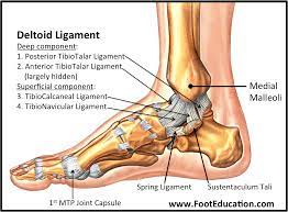
- Talus is geared to transfer forces from the foot through the ankle / talocruralleden to the bone.
- Calcanious is the largest bone of the foot. The Achilles tendon attaches on to the rear side of the calcaneus. On the front part attaches the plantar.
- Naviculare lies in front / anterior to the talus and the medial / inward to Cuboideum.
- Cuboideum leader of the calcaneus proximally, as well as with the fourth and fifth metatarsals distally and with the lateral Cuneiforme medial / inward.
- Each of Cuneiformebenen is wedge shaped. The medial, internal medial and lateral Cuneiforme forming point of the first three metatarsal distal and proximal radius bone. Cuboideum articulates with the lateral Cuneiforme.
- The five metatarsals articulate with the proximal phalanges.
- The big toe consists of two phalanges, three for each smaller toe.
- Although there are variations in the number and location of the sesamoids, it is usually two fixed sesamoid bone below the metatarsal head.
The tarsus
It is a complex bone arranged in two rows: the proximal and distal. The front or distal row consists of the medial and lateral sections. The lateral section is only one cuboid. In connection with the vertical stop position of the body bears the weight of the overlying card, which leads to the special structure of the tarsal bones in humans compared to animals.
It consists of three main elements:
- tarsus, whose bones include:
- calcaneus, which makes up the heel,
- astragalus on which support the leg bone (tibia, fibula)
- cuneiform bones,
- And the scaphoid.
- metatarsus (compound 5 bone)
- Phalanges: toe bones.
Astragals:
It is a cuboid bone interposed between the leg bones, calcaneus and the navicular. They distinguish a body, a head and a neck, and six faces. The upper face is entirely occupied by a trochlea, the lower face is divided by means of two facet joints with the heel, and the latter is mutually divided by the groove of the talus which, along with a shower present on the calcaneus, forms the sinus tarsi. Of these, two facets, the anteromedial is in turn divided into a front facet and a medium.
The cuboid:
It is a bone irregularly cubical which was before the heel, lateral to the scaphoid.Its upper face is wrinkled and not joint, the plantar has a marked ridge for the attachment of the plantar ligament long.
The side face is concave to allow passage of the peroneus longus tendon, the medial one, more extensive, offers the articular facet for the 3rd cuneiform. The rear surface of the cuboid bone articulates with the homologous face of the calcaneus. Even the front surface, split into two facets, it articulates with the bases of the 4th and 5th metacarpal.
Facts You Must Understand About The structure of Anatomy of The Foot.
Scaphoid
It is a bone-shaped spacecraft, anterior to the head of the talus, in contact with the proximal face of the three cuneiform. Of the two faces, the rear is provided with a glenoid cavity that receives the talar head, the front one, has three facet joints for many cuneiform. The medial end is provided with a big process marrow, the tuberosity of the scaphoid, the main point of insertion of the tendon of the tibialis posterior.
Cuneiform
There are three bones in the shape of triangular prisms, along their perimeter are provided with various planar facet joints, destined to the articulation with the cuboid, with the navicular, and with the first four metatarsal bones. The first cuneiform is the most voluminous; the second is distinguished to be the shortest.
Metatarsal bones
Five small long bones, placed between the tarsal bones and proximal phalanges. Each metatarsal bone has a body and two ends. The body has a triangular prismatic shape and describes an arc to the lower concavity. The proximal extremities have facet joints to the bones of the tarsus and metatarsals nearby, the distal articular surfaces are convex, welcomed the glenoid cavity of the proximal phalanges.
Metatarsals are 5, the first is as short and robust, and has a single facet joint and, on its surface takes plantar tendon insertion of the peroneus longus. Inferolateral corner presents the tuberosity of the 1st metatarsal, which is also the point of attachment to the peroneus longus.
The second metatarsal has the proximal end wedged between the three cuneiform, the third articulates with the lateral cuneiform and with 4 and 2nd metatarsal. The quarter is characterized by the joint surface in contact with the cuboid that is quadrilateral. The fifth metatarsal is the thinnest and its proximal end, presents the tuberosity of the 5th metatarsal, insertion of the tendon of peroneus short.
Phalanges
Long bones are small, less developed, although homologous to those of the hand.
Ankles: agility guarantees
The various joints divide the different tasks:
- Ankle joint: locomotion thanks to bending and stretching motion combined with a slight rotational component.
- Subtalar joint: uneven floors by a complex tilting rotary motion.
- Hock joints: spiral screw motion of front and hind foot, arch and stability.
- Metatarsophalangeal joints: shock absorbing roll, and repel.
Dynamics and Stability
- Stability: hind foot
- Agility: metatarsal
- Rolling: forefoot
- Shock absorption: sole
Foot muscles:
- Four muscle groups ensure the active interaction in the foot: Put, shock absorbing roll, and repel.
- The strong calf muscles ensure the necessary drive during braking and pushing off.
- The shin muscles surround the metatarsal as a stirrup. Rotate the rear foot outwards. The fibula muscles rotate the forefoot inward and so support the spiral.
- The short plantar muscle gives the arch support and resilience. You can be overstretched (flatfoot) or shortened (cavus).
Foot structure with brains
- Inside: vault
- Outside: ground contact
- Longitudinal arch: Stability
- Transverse arch: Elasticity.
In conclusion, the foot is a complex and vital structure that enables us to navigate the world with ease. Understanding the anatomy of the foot can help us appreciate its incredible design and take better care of our feet. So next time you take a step, remember the intricate workings of your foot that make it all possible.