Anatomy of Eyes information is very important for every medical student. Among all the senses eye has a special place. Assuming 100% information you perceive all the senses, taken together, that the share of 80% will have to the information received by the body from the outside. This is particularly important in the learning process. The eye reacts to light stimuli, which are electromagnetic waves in the visible spectrum. The human eye is a complex system. It is a complicated optical system.
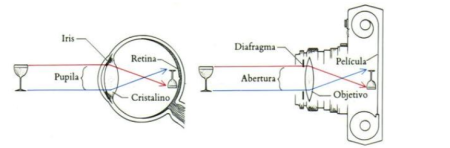
Light rays enter from the surroundings into the eye through the cornea. In order to understand how the eye, look at its structure. The eye is often compared to a camera, which has a casing (cornea), the lens (the lens), the aperture (iris) and a photosensitive film (retina). Furthermore it would be appropriate to compare the human eye with an analog cable complex computer device, as we look eye and see the brain.
Four parts of eyes:1) Peripheral or receiving part comprising:
eyeball
Protective device of the eyeball (the upper and lower eyelids, eye socket)
Eye adnexa (lacrimal gland, its ducts, conjunctivitis)
Oculomotor apparatus consisting of muscles.
2) Pathways – the optic nerve, optic chiasm and optic tract
3) sub cortical centers.
4 higher visual centers in the occipital lobes of the cerebral cortex
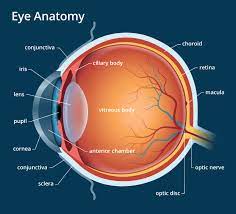
EYEBALL.
The eye is located in the orbit and is surrounded by soft tissue (adipose tissue, muscle, nerves, etc.). In front it is covered by the conjunctiva and eyelids covered. The eyeball is composed of three layers, limiting the interior space to the front, the rear chamber of the eye, as well as a space filled with the vitreous body – vitreous chamber.
Outdoor (fibrous) of the eye Submitted by dense connective tissue:
It consists of a transparent cornea anterior eye white and opaque sclera at the other extent. With elastic properties, these two shells form a characteristic shape of the eye.
What Is anatomy of the eye and How Does It Work?
Cornea:
The front of the eye is transparent thickness of about 500 microns in the central part. It accounts for approximately 60% of the refractive power of the eye. It has no blood vessels, and responds well to surgical simulation. The cornea has five layers: This transparent part (1/5) of the fibrous casing. Place her transition into the sclera is called limb. Ellipsoidal shape of the cornea, the vertical diameter – 11mm, horizontal – 12 mm. Corneal thickness of about 1mm. Transparency of the cornea due to the unique structure of it, in all of its cells are arranged in a strict order optical and lacks blood vessels.
The cornea is composed of 5 layers:
1.peredny epithelium
2.boumenova shell;
3.stroma;
4. Descemet’s hull;
5 .Back epithelium (endothelium)
The cornea is rich in nerve endings, so it is very sensitive. The cornea, not only misses but refracts the light rays, it has a large refractive power.
Endothelial cells:
A thin layer of cells in one row. This is Responsible for the power and transparency of the cornea. The cornea of the eye is very convenient object for refractive surgery because of its accessibility; its curvature can be modeled without opening the eyeball. In addition, avascular tissue capable of regeneration, allowing you to perform operations while maintaining its transparency. Finally, the cornea has a high refractive power that can significantly change it, even with minimal impacts.
Sclera
it is opaque portion of the fibrous sheath which has a white color. Despite its thickness of 1 mm, it is very dense and durable. The sclera is composed mainly of dense fibers, which give it strength. By the sclera of the eye muscles are attached.
Choroid
This is the average of the eye, which consists mainly of vessels of various calibers. Lines the posterior portion of the sclera, the retina adjacent to it, with which it is closely related. The choroid is responsible for blood supply of intraocular structures. In diseases of the retina it is very often involved in pathological process. The choroid is no nerve endings, so when her disease does not occur in pain, usually signaling on any malfunctions.
Iris
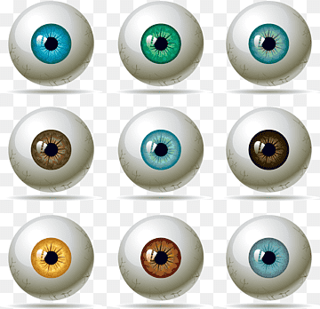
Shaped like a circle with an aperture inside (pupil). The iris consists of 2 muscles: narrows and widens the pupil contraction and relaxation during which the sizes of the pupil are changing. The structure of the iris consists of pigmented cells that determine eye color (if it is blue – so there’s little pigment cells, if the brown – a lot). Iris functions as a regulator of the width of the light beam entering into the eye.
Pupil
This is the hole in the iris. Its size depends on the level of illumination. The lighter, the pupil is less and vice versa. The average pupil diameter is 3-4 mm.
Internal retina (retina)
The retina is the first department of the visual analyzer. In the retina, the light is converted into nerve impulses, which are transmitted along the nerve to the brain, where these are analyzed, and the person perceives the image. The retina consists of 6 layers. The outer layer of the retina is the pigment. It absorbs light, reducing its dispersion within the eye. The following processes are layer of retinal cells, rods and cones. The optically active part of the retina can be seen when examining the eye. It is called the funds. In the funds can be considered vessels, optic disc (the place of exit of the optic nerve of the eye), as well as the macula. Macula – a region of the retina, where it is concentrated the maximum number of cones are responsible for color vision.
How does the human eye work?
When light reaches the eye, it will first pass through the cornea. This has a curved shape to be able to deflect the rays, allowing the light to pass through from the pupil to reach the lens.
On the other hand, there is an element that is in charge of regulating the light that enters, the iris (which we can understand as the colored part of our eyes). Well, in addition to giving it that characteristic color, it is also capable of regulating the amount of light that enters at all times, in combination with the ciliary muscles.
All this set will make the pupil contract when the light is intense, or dilate when there is little.
At the moment in which a light reaches the curve of the crystalline lens, it will reflect and be directed towards the retina. It will be at that exact point that the conversion of light into electrical energy will occur (something that the retina will do).
Finally, this energy will circulate through the optic nerve until it reaches the brain stem; when it is there, it will finally transform into the image we see, thanks to the help of the occipital lobe.
The human eye works like a camera.
The parts of the human eye are classically compared to a camera, made up of focusing lenses and an iris as a diaphragm, which can be opened or closed to control the amount of light that passes into an area at the back of the eye. which we call the retina.