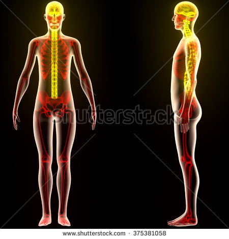
Human skeleton is the bony framework of the body.The Knowledge of human Skeleton System is very valuable for every science student. Its functions are twofold: (a) blood formation—the cells of the blood are derived from the cancellous tissue and marrow of bone; (b) it gives definite shape and rigidity to the body and forms a supporting framework which surrounds and protects the most delicate and important viscera. Its rigidity supplies fixed points from which the muscles act in moving the limbs and trunk.
For purposes of description, the skeleton is divided into (i) the axial skeleton, consisting of the skull, vertebral column, hyoid bone, sternum, and ribs; (2) the appendicular skeleton, comprising the limbs.
The adult skeleton is formed of two hundred bones.. These are classified as follows:
Long bones—e.g. those of the limbs.
Short bones—e.g. mist (carpus) and ankle (tarsus).
Flat bones—e.g. skull.
Irregular bones—e.g. vertebral column.
Sesamoid bones are developed in tendons; the patella is the most important.Long bones consist of a shaft and two extremities; their structure.Short and irregular bones are composed of cancellous tissue with a thin covering layer of compact tissue.Flat bones consist of two layers of compact tissue, between which a layer of cancellous tissue, the dipole, is enclosed.All bones show markings upon their external surface, due to pull of muscles, or the forming of channels for blood vessels and nerves. These markings are in the form of Projections or depressions.
Bones of the Upper Extremities
Sixty-four bones form the shoulder girdle and upper extremities.
The shoulder girdle is formed by the two scapulae and clavicles. It is completed in front by the upper end of the sternum; behind it is imperfect, being connected to the trunk by muscles only.
The clavicle or collar-bone forms the front part of the shoulder girdle. It is a long bone, the shaft of which presents a double curve, with its convexity forwards in the inner two-thirds where the bone is rounded, and backwards in the outer third where the bone is flattened.
Articulations. The clavicle articulates with the sternum and with the acromion process of the scapula.
The scapula or shoulder-blade is a flat bone. It has two surfaces, anterior and dorsal; three margins, superior, axillary, and vertebral; and three angles, internal, external, and inferior.
The anterior surface is concave and forms the subscapular fossa. The dorsal surface is unequally divided by a ridge, the spine of the scapula, which terminates in a triangular projection, the acromion process; this ridge divides the supraspinous fossa above from the infraspinous fossa below. In the external angle is a hollow, the glenoid cavity, on to which the head of the humerus fits. Below the glenoid cavity is a constriction known as the neck, and jutting forwards from the neck is the coracoid process.
Articulations. The scapula articulates with the clavicle and humerus.The humerus, radius, and ulna are long bones, each having a shaft and two extremities.
The humerus. The upper extremity of the humerus consists of a head, an anatomical neck, and a greater and lesser tuberosity divided by a groove — the bicipital groove — in which plays the long tendon of the biceps muscle. The head is rounded, smooth, and covered by hyaline cartilage.
What Everyone Ought To Know About Human Skeleton.
The anatomical neck is a constriction separating the head from the tuberositiesties. The surgical neck is
the narrower part of the bone below the tuberosities tiesties, so called because it is commonly the part to fracture. The shaft is cylindrical and becomes flattened towards its lower end. About the middle of its external surface is a rough elevation, the deltoid tubercle (or insertion tiontion of deltoid), and about half-way down a shallow groove
crosses the bone obliquely downwards, and behind—the musculo-spiral groove, along which passes the musculo-
spiral (radial) nerve.
The lower extremity presents two articular surfaces” joined by a shallow groove; the outer is the capitellum and articulates with the radius’, the inner or trochlear surface fits into the greater sigmoid cavity of the ulna. On either side of these surfaces are the external epicondyle, from which the extensor muscles of the forearm take origin, and the internal epicondyle, from which the flexor muscles arise; the internal being larger than the external.
Above the trochlear surface and the capitellum in front are two depressions—the coronoid and radial fossae, which receive the coronoid process of the ulna and the head of the radius respectively, when the forearm is flexed; at the back is a deep depression, the olecranon fossa, into which fits the olecranon process of the ulna in extension.
Articulations. The humerus articulates with the scapula, ulna, and radius.The ulna is the inner bone (little finger side) of the forearm. The upper extremity of the ulna is thick and strong and has two processes—the olecranon behind, which .forms the point of the elbow, and the coronoid in front.
The greater sigmoid cavity is the large notch between these processes; it receives the trochlear surface of the humerus. The lesser sigmoid cavity is a notch on the outer side of the coronoid process for articulation with the head of the radius.
The shaft tapers gradually from above downwards. The lower extremity consists of a round head from which projects the styloid process. The distal surface of the head rests on a pad of cartilage, the triangular fibro-cartilage of the wrist-joint, but does not articulate with the wrist- bones.
Articulations. The ulna articulates with the humerus and radius.
The radius is the outer bone (thumb side) of the forearm. The upper extremity consists of a head, a neck, and a bicipital tuberosity. The head has a concave upper surface for articulation with the capitellum of the humerus, and on its inner side is an articular surface, for articulation with the lesser sigmoid cavity of the ulna, within which it rotates in the movements of pronation and supination.
The neck is the constricted portion below the head.The bicipital tuberosity lies below the neck, on the inner side of the bone; into it is inserted the biceps muscle.The shaft is narrower above than below and slightly curved.The lower extremity has two articular surfaces, the ulnar facet on the inner side, which receives the head of the ulna, and an articular surface on the distal aspect which articulates with the carpal bones. The styloid projects downward from the inner and back part of the bone.
Articulations. The radius articulates with the n i, humerus, ulna, and wrist bones.
Bones of the wrist or carpus.
The carpal bones are short bones, eight in number and arranged in two rows.The upper row from radial to ulnar side are named:Scaphoid (navicular), boat-shaped. Semilunar (lunate), half-moon shaped. Cuneiform (three-cornered bone), wedge-shaped. Pisiform, shaped like a pea.
The lower row in the same order are named:
Trapezium (large multangular).
Trapezoid (small multangular).
Os magnum (capitate).
Of these the scaphoid and semilunar. form a condyle and articulate with the radius to form the wrist-joint.The metacarpal bones are five in number and form the skeleton of the palm (metacarpus).The carpal extremity, or base, of each articulates with one or more carpal bones; the shafts are separated by the interosseous spaces. The digital extremity, or head of each, articulates with the upper proximal phalanx of the corresponding finger.The phalanges are the finger bones. They are long bones and number fourteen, three for each finger and two for the thumb.
The pelvic girdle is a bony ring formed by the sacrum and coccyx behind and the two hip bones (innominate bones) in front.The os innominatum, or hip bone, is a flat bone, irregular in shape. In a young child it consists of three bones, the ilium, ischium and pubis, but as growth proceeds these become united.
The acetabulum is a deep depression, or cup-shaped cavity, on the outer surface of the bone; it is formed by portions of the three bones and receives the head of the femur; it has a prominent margin broken by a notch. Continuous with this notch is a rough depression, in which is a pad of fat. Around this is the articular surface and attached to the margins of the notch is ligamentum teres.
The obturator foramen is the large opening which lies below the acetabulum. The obturator membrane stretches across the opening from edge to edge and forms a canal for the obturator vessels and nerve on their passage to the thigh.
The great sciatic notch lies below the posterior part of the ilium. It is converted by a ligament into two fora- tnina, through which pass the great sciatic nerve to the thigh, the gluteal vessels and nerve to the buttocks and the pyriformis muscle.
The ilium is the large expanded portion of bone above the acetabulum; it has a crest, an internal and external surface, and an anterior and posterior border. The crest terminates at either end in the anterior and posterior superior iliac spines.
The external surface is divided into a lower acetabular and an upper or gluteal portion. The upper portion is smooth and its surface is crossed by three curved lines— superior, middle, and inferior, for attachment of the gluteal muscles.
The internal surface presents a smooth concave surface, the iliac fossa; behind this is a rough surface, auricular surface (like an ear), which articulates with the sacrum. Below is a smooth, rounded border, the ilio-pectineal line, separating the false from the true pelvis.
The ischium is the lower and back part of the hip bone; it consists of a body, a tuberosity, and a ramus. The body forms part of the acetabulum. The tuberosity is that part of the bone on which the body rests in sitting. The ramus extends upwards from the tuberosity and unites with the ramus of the pubis.
The pubis is the front part of the hip bone; it has a body, an ascending and a descending ramus. The body is the broadest part where the rami meet. The ascending ramus extends from the body to the acetabulum.
The descending ramus extends downwards to unite with the ramus of ischium. The os pubis articulates with its fellow of the opposite side at the symphysis pubis. The 0s innominatum articulates with its fellow, with the sacrum, and the femur.The femur, tibia, and fibula are long bones each having a shaft and two extremities.
The femur, or thigh bone, is the largest and longest bone in the body. Its upper extremity has a head, a neck, and two trochanters; the head is rounded and near its centre is a fossa called the fovea for the attachment of the ligamentum teres.The Neck connects the head to the shaft,which joins at an angle.
The greater trochanter is a square mass of bone, which projects upwards at the junction of the shaft and the neck. The lesser trochanter is a cone-shaped process situated also at the juncture of the shaft and neck. It projects inwards and backwards The shaft is bent forward slightly; it is smooth except for a rough line, the linea aspera, down the back for the attachment of muscles.
The lower extremity is broad and divided into two condyles—thick, rounded masses of bone which project backwards beyond the surface of the shaft. Between the condyles, is the intercon- dyloid or popliteal notch.In front, between the condyles, is the smooth patellar surface on which the patella rests.
Articulations. The femur articulates with the innominate bone, the patella, and the tibia.
The patella is a sesamoid bone, an ossified part of the extensor tendon; it forms the knee-cap.
The tibia is the inner bone of the leg. The tipper extremity, or head, consists of two tuberosities, internal and external, each having a smooth surface above for articulation with the corresponding condyle of the femur. A ridge of bone, the spine articular surfaces. Below the front of the head of the tibia is a rounded projection for attachment of the ligaments of the patella, the tubercle of the tibia. The shaft is triangular in shape; its inner surface lies beneath the skin, its outer surface is covered by the anterior muscles between the tibia and fibula.
The lower extremity enters into the formation of the ankle joint. It presents an articular surface below for the astragalus, and is prolonged downwards on the inner side to form the internal malleolus, or ankle bone, and on the outer side has a facet for articulation with the fibula.
Articulations. The tibia articulates with the femur, the fibula, and the astragalus.
The fibula is the outer bone of the leg. Its upper extremity, or head, lies behind the outer tibial tuberosity, with which it articulates. The shaft is irregular and lies deeply buried among the leg muscles. The lower extremity lies just beneath the skin, forming the external malleolus, which projects further down than the internal. An articular surface for the astragalus faces towards the ankle joint; behind this is a rough, deep depression, to which is attached a strong band of the external lateral ligament of the ankle joint.
Articulations. The fibula articulates with the tibia and astragalus.
Every Anatomy Student Must Know The Ultimate System Of Human Skeleton
The tarsus, or ankle, is composed of seven bones:
- The astragalus (talus) consists of a body, a neck, and a head. The body fits between the two malleoli to form the ankle joint. The head projects forwards to articulate with the scaphoid. The lower surface has a double facet for articulation with the os calcis.
- The os calcis (calcaneus) is the largest bone of the tarsus: the back part projects behind the astragalus and forms the part of the heel which rests on the ground, the front part articulates with the cuboid. A ridge of bone, the sustentaculum tali, projects forward for support of the astragalus.
- The scaphoid (navicular) lies on the inner margin of the foot between the astragalus at the back and the three cuneiforms in front.
- The cuboid lies on the outer side of the foot between the os calcis and the two outer metatarsals.
- The cuneiform bones are three wedge-shaped bones lying side by side between the scaphoid and the three inner metatarsals.
- The metatarsal bones are five in number. Each bone articulates by its proximal extremity with one or more of the tarsal bones and its neighbouring metatarsal, and by its distal extremity with the first row of phalanges. .
- The phalanges (singular, phalanx) number fourteen —two for the great toe and three each for the other four toes.
The Spinal Column
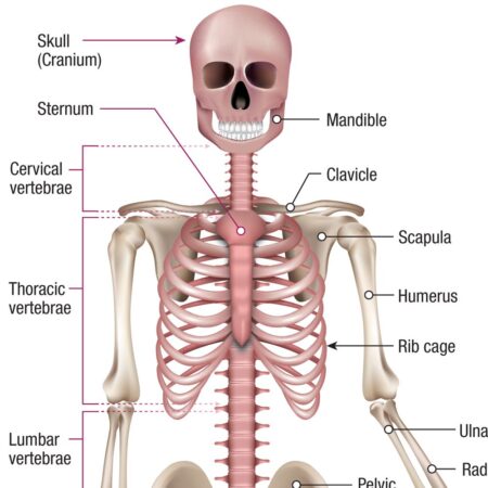
The spinal column is composed of thirty-three bones called vertebrae, named according to the region in which they are found:
- Cervical vertebrae of neck, 7.
- Dorsal vertebrae of thorax, 12 Lumbar vertebrae of loin, 5.
- Sacral vertebrae of pelvis, 5 joined as one.
- Coccygeal vertebrae of tail bone, 4 joined as one.
The vertebrae differ in size and shape, but a general plan of structure is typical of all. A typical vertebra is composed of the body, a central mass of cancellous bone that forms the anterior portion, and gives the main support. The pedicles are two processes of bone extending backwards to join with the laminae in the formation of the neural arch or spinal canal. At the junction of the laminae and pedicles, there pass off, at each side, the transverse processes: and where the laminae meet behind is given off the spinous process of the vertebra.
The meeting place of the transverse process, lamina, and pedicle is marked on each side by two articular processes, making four for each vertebra. These are the superior and inferior articular processes. The inferior ones of the vertebra above articulate with the superior ones of the vertebra below. Above and below each pedicle are the intervertebral foramina, through which the nerves leave the spinal cord. The spinal canal formed by the arches of the vertebrae contains the spinal cord.
Characters of the vertebrae in different regions.
Cervical vertebrae. The bodies are small and the neural arch large.The spinous processes are short and forked at the end. The transverse processes contain a foramen which transmits the vertebral artery. Peculiar cervical vertebrae. The first cervical, the atlas, so called because it supports the head, has no body or spine: it consists of an anterior and a posterior arch, and two lateral masses each presenting an articular facet above and below.
The two facets on its upper aspect receive the condyles of the occipital bone, forming the joint through which the nodding movement of the head takes place. The second cervical, or axis, has projecting upwards from the body a process,the odontoid process from the detached body of the atlas,this passes through the ring of the atlas,forming the pivot on which the head rotates.
The sacrum is a triangular bone formed by five vertebrae fused together. The transverse processes are fused to form the lateral mass, at the upper and outer sides of which are the auricular surfaces which articulate with the ilium, forming the sacro-iliac joint. Four pairs of foramina in the front, and four at the back (8 pairs), pierce the bone. The coccyx consists of two to four bones fused to form one, it articulates with the lower end of sacrum,
Intervertebral discs. Between bodies of the vertebrae are elastic discs of fibrocartilage which form elastic pads between the vertebrae that prevent jarring of the spine. The joints between the discs and vertebrae are each only slightly movable, but the flexibility of the column as a whole is considerable.
The curves of the spine.
- The cervical curve which has its convexity forwards.
- The thoracic curve which has its concavity forwards.
- The lumbar curve which has its convexity forwards.
- The pelvic curve which has its concavity forwards.
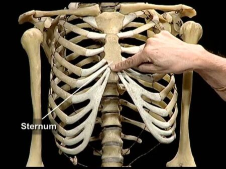
Bones of the Chest or Thorax
The spine of the thorax consists of twelve dorsal vertebrae as described. There are twelve pairs of ribs, one pair to each dorsal vertebra.The upper seven ribs are joined directly to the sternum,It is long and narrow and notched for the attachment of the costal cartilages.
The Skull
The skull includes the cranium and the face.
Eight bones form the cranium:
Frontal, i. Parietal, 2. Occipital, 1. Temporal, 2.
Ethmoid, 1. Sphenoid, 1.
The frontal bone consists of a frontal part—the forehead •—{wo orbital plates, and a nasal part. The frontal part unites above with the parietal bones: below it is bounded by the supraorbital margins with the nasal part between. Above the supraorbital margins are the superciliary ridges, to which muscles are attached. The frontal sinuses are two air-containing cavities in the bone which communicate with the nasal cavity.
The parietal bones help to form the roof and sides of the skull.
The occipital bone is at the back and base of the skull. The back part is joined to the parietal bones; its most prominent point is the occipital protuberance. The basal portion is pierced by the foramen magnum, which transmits the spinal cord and vertebral artery. At the sides of the foramen magnum arc the two condyles which rest on the atlas.
The temporal bones are one on either side: each consists of a squamous, a petrous, and a mastoid portion.The squamous portion fills the gap between parietal, occipital, and sphenoid bones: from it projects the zygomatic process to join the malar bone; beneath the beginning of this is the external auditory meatus, just in front of which the mandibular fossa receives the condyle of the lower jaw bone.
The petrous portion—a hard mass projecting inwards contains organs of hearing and balance: the styloid process projects from its lower surface. The mastoid process forms the prominence behind the ear: it contains a cavity—the mastoid antrum, and smaller cells which communicate with the cavity of the ear The ethmoid is an irregular mass of bone wedged between the orbits: it comprises two lateral masses, a horizontal— cribriform—plate, perforated for the passage of the olfactory nerves from the nose, and a perpendicular plate which forms the upper part of the nasal system.
The sphenoid bone lies immediately behind the ethmoid: its shape resembles a bat with wings spread, it consists of a body, wings, and two pterygoid processes: its body presents a saddle-like depression, the sella turcica (Turkish saddle) or pituitary fossa, in which the pituitary gland rests; it contains one or more large air cells, the sphenoidal sinuses, which open into the nose.
Bones of the face
Nasal, 2. Lachrymal, 2. Malar, 2. Palate, 2. Inferior turbinate, 2. Vomer, 1. Inferior maxillary or mandible, 1. Superior maxillary, 2 (united form maxilla).
The nasal bones form the bridge of the nose.
The lachrymal bones, situated in the walls of the orbits, contain the beginnings of the canals in which the lachrymal (tear) ducts run.
The malar, or cheek bones, form the prominence of the cheeks.
The maxilla is composed of the two superior maxillary bones united: it presents a body and several processes; the body is hollow, the space being called the maxillary sinus or antrum of Highmore. The alveolar process is that part of the bone in which the upper teeth are fixed.
The palate process forms part of the hard palate. The two palate bones complete the hard palate.The inferior turbinated bones are situated on the outer walls of the nasal cavity.
The vomer forms the lower part of the nasal septum.
The mandible (lower jaw bone) is the only moveable bone of the face: it consists of a horizontal part, the body, which contains the lower teeth, and two vertical rami (branches) which join it at an angle—the angle of jaw. The rami each present two processes, the condylar process which articulates with the mandibular fossa of the temporal bone, and the coronoid process.
The skull as a whole is a somewhat globular case for the brain, the cranium, with the irregular face bones attached. The upper surface of the cranium is known as the vault,and the lower surface as the base of the skull: its outer surface is marked by the sutures where the bones unite.The coronary suture runs from one temple to the other between the frontal and parietal bones.The sagittal suture is in the midline between the two parietal bones.
The lambdoidal suture is A shaped, and lies between the parietal bones and the occipital bone.
Fossae of the skull.
The inner surface of the cranium is modelled to fit the brain, the bones are marked by the convolutions of the brain and by large veins and arteries. The interior of the base of the skull is divided by two pairs of radiating ridges into the three cranial fossae.
The anterior fossa lies in front of the small wings of the sphenoid bone and supports the frontal lobes of the brain.
The middle fossa lies behind the small wings of the sphenoid and in front of the petrous part of the temporal bone: its lateral parts lodge the temporo-sphenoidal lobes of the brain.
The posterior fossa surrounds the foramen magnum: it lodges the cerebellum and medulla.
The groove for the lateral sinus, which is a large vein receiving the blood from the brain: it makes a deep groove round the edge of the posterior fossa, which underlies the mastoid process and extends to the jugular foramen.
The groove for the middle meningeal artery runs upwards and outwards across the middle cranial fossa.
The temporal fossa is the thinnest part of the skull: it lies above the zygomatic arch and is occupied by the temporal muscle.
The zygomatic fossa is below the zygomatic arch.The orbital fossa, or cavity for the eye, is pyramid shaped, the apex to the back: its roof is formed by the orbital plate of the frontal bone, its floor by the maxilla: the lacrimal and ethmoid bones are on the inner wall, the sphenoid and malar on the outer. The optic nerve enters at the apex.
The nasal fossae (nose) are two cavities separated by a septum partly composed by the ethmoid and vomer and completed by cartilage. The roof is formed by the nasal, ethmoid, and frontal bones, the floor by the maxillary and ethmoid bones. The anterior nares are the openings on to the face: the posterior nares open into the naso-pharynx. Turbinated bones are seen in the outer walls.
Foramina.
The foramen magnum in the occipital bone transmits the spinal cord and its membranes, the vertebral arteries, and spinal accessory nerves.At the base of the skull the foramen lacerum transmits the internal carotid artery.
The jugular foramen transmits the jugular vein and the pneumogastric and glossopharyngeal nerves.
The foramen rotundum transmits the second, and the or breast bone, by the costal cartilage. The next three ribs are joined by cartilage to the rib above and so indirectly to the sternum. The last two ribs are the ‘ floating ribs.’ In front they are free and unconnected with any bone. Each rib is a long bone which has a rounded head that