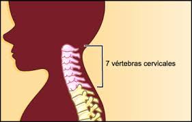
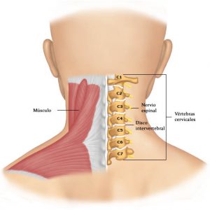
Cervical Region The spine is made up of 24 bones, called vertebrae. The first 7 vertebrae form the cervical spine. Technically we refer to them as vertebrae C1 to C7. The cervical spine begins where the upper vertebra (C1, atlas) connects to the base of the skull. The first vertebrae form the cervical spine. The cervical spine is the most mobile segment of the spine. It is formed by seven vertebrae and in addition to eight pairs of cervical nerves.
The second cervical vertebra is an axial vertebra ( epistrophy ). It is characterized by the presence of a tooth instead of the head and the crest instead of the spinous process. In ruminants, the tooth is in the form of a half-cylinder, in horses it is chisel-shaped, in pigs it is blunt, conical. In horses, the crest of the axial vertebra is bifurcated and fuses with the caudal articular processes. In a pig, the crest of the axial vertebra is narrow, high, directed caudally.
Characteristics Of The Cervical Vertebrae
The cervical vertebrae, which differ by presenting individual characteristics different from the general ones, are:
- the atlas: It is the first cervical vertebra (C1), its name refers to the mythological titan that held the celestial vault on its shoulders. It has no body or thorny process, but has an anterior and posterior arch, which are joined by the lateral masses. Each lateral mass has a concave and ellipsoid upper articular facet. This veneer is articulated with the occipital bone condyle. On the underside of each lateral mass there is a flat articular facet to articulate with the axis. The transverse process is born from the lateral face of the lateral mass, which is longer than that of the other cervical vertebrae. Both arches present in the midline, an anterior and a posterior tuber respectively, having a very large vertebral hole. It articulates with the tooth of the axis through an articular surface called dental fosita. The groove of the vertebral artery is seen on the upper side of the lateral end of the posterior arch. The first cervical nerve passes behind the lateral mass.
- the axis: it is the second cervical vertebra (C2) and its name means axis. It has a vertical eminence that is directed upward from the upper end of its body called the axis tooth (apophysis odontoid) and around it rotates the atlas next to the skull. It has an articular surface on its anterior face to articulate with the dental atlas of the atlas in the median atlantoaxil joint and on its posterior face, the posterior articular veneer, for the transverse ligament. On both sides of the tooth of the axis presents two articular surfaces to articulate with the atlas in the lateral atlantoaxial joints. They have two lower articular veneers, for the third cervical vertebra. The thorny process is wide and short.
- the sixth: The sixth cervical vertebra (C6), is characterized by the greater development of the anterior tubercle of its transverse process, which due to its importance for the compression of the common carotid artery during surgery, has been called a carotid tubercle.
- seventh vertebrae: the seventh cervical vertebra (C7), is a transition vertebra, and has some characteristics that resemble a thoracic vertebra. The end of its spiny process does not bifurcate, it is especially long and embossed under the skin, so it is called a prominent vertebra and easily palpated. Transverse processes are unituberculous, have no transverse hole or are very small.
Characteristics of the vertebrae typical of the cervical region
They are from the third to the sixth and have the following characteristics:
- The body is made up of very vascularized spongy tissue, covered by a thin layer of compact bone tissue. It is approximately cylindrical in shape, occupies the anterior part of the bone, is arranged vertically and its fundamental function is that of support. Its transverse diameter is larger than the anteroposterior diameter and has some distinguishing details. At the lateral ends of its upper face it has two small eminences, the unciform processes (hook-shaped), arranged as a semilunar elevation of the lateral edges. On the lateral ends of the lower face of the body there are two small recesses that serve for articulation with the unciform processes of the underlying vertebrae, giving this face a convex shape.
- Vertebral arch, the vertebral plates are flattened and quadrilateral, form most of the posterolateral wall of the foramen or vertebral hole. The medial and posterior end of each sheet is confused with the base of the spinous process (apophysis). They have a posterior face covered by the muscles (mm) deep in the back. In general they are arranged in an oblique plane down and back. It joins by its anterior ends to the posterolateral limits of the body. This arch is concave forward. The vertebral hole is triangular, large in relation to the body and with a wider anterior base, corresponding to the thickness of the spinal cord at this level. Its function is essentially protection.
- The vertebral pedicles are implanted in the vertebral body at a point closer to its upper face than to the lower one, they are two irregularly cylindrical, flattened, thin and narrow bony portions found at the anterior ends of the vertebral arch. They extend from the back and side of the vertebral body to the junction of the base of the transverse process with the two joint processes on each side. Each pedicle has two vertebral recesses, one at its upper edge and another at its lower edge, these recesses overlapping with those of the neighboring vertebrae form on each side of the spine a series of holes called intervertebral forams. The short distance between the body and the joint processes in the cervical region determines that at this level these holes are narrow.
Cervical region movements
The movements between two neighboring vertebrae are not wide. However, thanks to the large number of bone segments of which the spine is constituted, the small movements between isolated vertebrae, when added together, give it a wide mobility with the cervical region being the most mobile. The following movements are possible:
- In the frontal axis flexion and extension (ventral flexion and dorsal flexion).
- Sagittal axis right lateral flexion and left lateral flexion.
- With the combination of angular movements, around the frontal and sagittal axes, circumduction movements are performed.
Irrigation of the cervical spine
It is in charge of the posterior spinal arteries that penetrate the vertebral canal or spinal canal through the intervertebral hole, these branches come from the vertebral artery, the deep cervical and the anterior or ascending cervical (lower thyroid branch).
Venous Drainage
It is performed by veins that correspond to the aforementioned dorsoespinal arterial branches, accompany them in the intervertebral hole and form a true plexus around them. They flow into the vertebral veins.