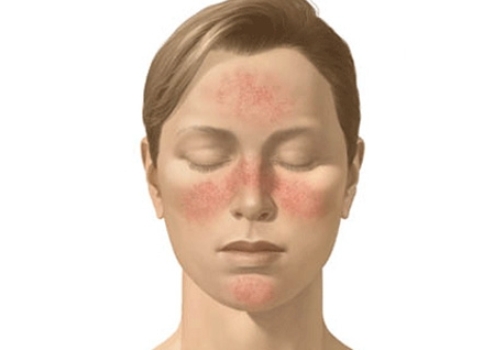
Systemic lupus erythematosos (SLE) is a chronic, inflammatory disease of unknown cause a fleeting skin, joints, kidneys, nervous system. serous membranes, and often other organs of the body. The classic facial “butterfly rash” facilitates diagnosis. although the rash need not be present. The clinical course may be fulminant or indolent, but generally is characterized by periods of remissions and relapses. Patients with SLE develop distinct immunologic abnormalities, especially antinuciear antibodies. A diagnosis of SLE hits frequently been established or confirmed by the finding of these antibodies, especially the one causing the LE cell phenomenon.

Incidence And Etiology of Systemic Lupus Erythematosos
SI.K is not a rare disorder. The prevalence rate is approximately 5 per 100,000 population The disease appears to occur more frequently in blacks than in whites and is rare among Asians. It occur.’ live to ten times more frequently among females than males and has been diagnosed in patients ranging from 2 to 97 years old However, the majority of patients are first discovered to have SLE while in their third and fourth decades of life.
The cause of SI.K remains unknown Exposure to sunlight or ultraviolet radiation, viz., sunlamps, often leads to the prompt appearance of the facial butterflv rash or a rash on other exposed skin surfaces. Many other factors, including infections, surgery, and certain drugs, have been shown to be associated with exacerbations of SLE Recent attention has focused on procainamide. hydralazine, hydantoins, and other drugs which may precipitate a lupus-like illness.
In view of the preponderance of the disease in females, endocrine factors have been thought to influence the development of SLE The disease tends to remit during the last two trimesters of pregnancy and to relapse post par-turn. Inhibitors of ovulation can cause an exacerbation of SLF
Genetic factor may influence the development of SI.K in Musccptihle individual’ Much evidence MUpports Uiik hypothesis The high incidence of SLE unions females might Im- considered an X-chromosome-related factor Connective tissue disorders, dyNgammaglohulinemia and autoimmune phenomena are obsem*d frequently among relatives of many patients with SLE. Genetic control of immune responses to certain antigens correlates closely with histocompatibility types in some unimnls. An association between particular HLA antigens and SLE has been noted. suggesting that SLE patients may hyperreact to some antigens Additional evidence for :i genetic role in SLE comes from studies in certain strums of inbreil mice and dogs NZB and .WAV mice develop an illness which includes hemolytic anemia.
Lupus erythematosus is characterized particularly by autoimmune phenomena. Patients develop antibodies to many of their own cells, cell constituents, and proteins. The origin of these autoantibodies is as obscure as is the cause of SLE. However, they do appear to represent a loss of tolerance to self antigens M ice of the NZB strain have been found to lose their tolerance for foreign antigens much more rapidly than do other mice.
Pathogenem and Pathology Many of the manifestations of SLE appear to result from the deposition of antigen-anti body complexes in tissues Although immune complexes have not been isolated from SLE patients, certain evidence is consistent with their presence. In laboratory animals, when immune complexes are formed in the circulation, serum complement levels fall and nephritis develops. In patients with SLE. especially in those with anti-DNA antibodies, a fall in complement levels is often associated with the development of nephritis. In addition, some patients with serum antibodies to DNA develop fever and nephritis as the antibody disappears and is replaced by free DNA. a sequence which presumably represents immune complex formation and deposition followed by antigen excess Furthermore. some sera from SLE patients with nephritis, when treated with deoxyribonuclease, have a n*e in anti-DNA antiliody levels, This fact suggest- that DNA has been hound in vivo to the antibody as an immune complex. Using the immunofluorescent technique, immunoglobulins (especially IgG), complement components, and antigens i viz.. DNA i have been detected in a granular and lumpy distribution along the renal glomerular basement membrane.
Diagnosis of Systemic Lupus Erythematosus;
The diagnosis of SLE can be made if any four or more of the following manifestations are present, serially or simultaneously, during any interval of observation butterfly rash, discoid lupus. Raynaud’s phenomenon. alopecia, photosensitivity, oral or nasopharyngeal ulceration, arthritis without deformity, LE cells, chronic false-positive STS. profuse proteinuria, cellular casts, pleuritisor pericarditis, psychosis and/or convulsions, or hemolytic anemia, leukopenia, or thrombocytopenia SLE should also be suspected especially in young females with unexplained fever, purpura, easy bruising, diffuse adenopathy, hepatosplenomegaly, peripheral neuropathy.endocarditis, myocarditis, interstitial pneumonitis. peritonitis, or aseptic meningitis.
Patients are frequently first diagnosed as having rheumatoid arthritis, rheumatic fever (especially children glomerulonephritis, tuberculosis, scleroderma, vasculitis <including periarteritis), idiopathic thrombocytopenic purpura, lymphoma, anemia, or neutropenia. The LEcell is specific for SLE. if rheumatoid arthrit is, lupoid hepatitis, or drug reactions can be excluded. Although the LE cell test is quite often negative, the antinuclear factor test is positive in virtually all patients with SLE. The antinuclear antibodies have also been detected in K percent of patients with Sjogren’s syndrome. -Id to 75 per cent of patients with scleroderma.
Diagnostic problems always arise in patients with the so-called overlap syndromes, mixtures of signs and symptoms of SLE und other related diseases, such as rheumatoid arthritis, Sjogren’s syndrome scleroderma, and dermatomyositis. These patients are best considered. and treated, for their primary symptom- and complaints.
Therapy and Treatment OF Systemic Lupus Erythematosus;
Ac there are no specific remedies for the treatment of the underlying processes of SLE. the main goals of management must In- limited to the treatment of acute relapses and prevention of exacerbations. Certain measures are helpful in prevention of relapses. Sunlight, ultraviolet radiation, immunizations, blood transfusions, penicillin, and sulfonamides should be avoided when possible. Surgery or infections max lead to a relapse and may require more aggressive concurrent management of the lupus. In the long-term management of this illness, both patient and physician must learn to recognize those symptoms and signs that herald relapses Although these symptoms may involve fever and arthralgia in some patients, they may appear as hair loss, mucosal ulcers, pleurisy, weight lo>-. or simply fatigue in others. Before frank clinical symptoms develop, many patients will develop abnormal laboratory tests such as a reduced level of hemoglobin or serum complement or a reduced white blood cell or platelet count, an abnormal urinalysis or creatinine clearance.
Active disease should be treated aggressively to prevent permanent tissue injury. The general measures to be taken include rest when the disease is active, sunscreens, and physical therapy for muscle weakness and deformities. For those patients on moderate to high dosages of corticosteroids, salt restriction and diuretics may he of benefit. Drugs used for the treatment of SI.K include topicul steroids, salicylates, anlima-lariuls, corticosteroids, and immunosuppressive*. The use of corticosteroids is indicated in most patients with SI.K and has improved survival. Dosage should be individualized according to the activity of the disease and side effect* ImmunonuppregHiven are best used according to the guidelines of Schwartz and Cowans, whose indications are life-threatening or seriously crippling disease, presence of reversible lesions; failure to respond to conventional therapy or intolerable side effect*; no active infection; no hematologic contraindication; meticulous follow-up, and objective evaluation.
The acute erythematous maculopapular rash responds well to the avoidance of sunlight, local application of corticosteroid cream, and the systemic introduction of an-timalarials such as hydroxychloroquin. Systemic corticosteroids are rarely warranted The chronic lesion, or discoid lupus, responds to these same measures to a variable degree. Severe, extensive lesions have been treated with large doses of antimalarials when all else has failed. However, the patient should he cautioned about the relative risks of retinal damage when taking large doses of antimalarials. Arthralgia and arthritis respond well to rest, physical therapy, splinting, salicylates, and antimalarials. Steroids should not be needed.