The Skeleton and the movements, Why we need a skeleton, Bones and muscles work together, The bone structure, Cartilage, ligaments and tendons, The bones of the head, The spine, The rib cage, The bones of the arms and legs , Joints.
Bone functions
Bones have the following functions:
- Support: they serve to support the different parts of the body;
- Movement: they serve as an insertion point for the muscles, thus helping with movement;
- Protection: some bones protect important structures, such as the cranial box, which protects the brain;
- Reserve of mineral salts: bones store various salts, such as calcium, phosphorus, potassium, among others. In relation to calcium, when the amount eliminated by the body is greater than that absorbed, the body seeks balance by removing calcium from the bones.
→ Classification of bones
There are several ways to classify bones. The most used and widespread takes into account its dimensions :
- Long: have a length greater than width and thickness. Examples: Femur and tibia.
- Short: have equivalent length, width and thickness. Examples: Carpal and tarsal bones.
- Laminar: they have the equivalent width and length, and greater than the thickness. Examples: Scapula and hip bones.
The characteristics of some bones do not, however, allow them to fall into this classification. Therefore, they are present in the following categories :
- Irregular: they have dimensions and shapes that do not fit the previous classification. Example: Vertebrae.
- Pneumatics: have one or more cavities filled with air. Examples: Frontal and maxillary.
- Sesamoid: are bones surrounded by tendons and fibers. Example: Patella.
What are bones for?
Bones perform several functions:
- They act as a reserve and storage of minerals such as calcium, phosphorus, sodium and magnesium, essential for regulating many physiological mechanisms.
- The bone marrow in them is capable of producing blood cells (white blood cells, red blood cells, platelets).
- They give shape and support to the parts of the body from which they are made.
- Thanks to the interaction with the muscles and tendons they allow the movement of the limbs.
- They have important protective functions (think of the importance of the skull in protecting the brain).
- They provide support and stability even in the absence of movement.
Bone Structure
The bone structure consists of several types of connective tissue (dense, bony, adipose, cartilaginous and blood), in addition to nervous tissue.
The long bones are formed by several layers, see the table below:
| Bone layer | description |
|---|---|
| Periosteum | It is the most external, being a thin and fibrous membrane (dense connective tissue) that surrounds the bone, except in the articulation regions (epiphyses). It is in the periosteum that the muscles and tendons are inserted. |
| Compact bone | The compact bone tissue is composed of calcium, phosphorus and collagen fibers that give it resistance. It is the most rigid part of the bone, formed by small channels that circulate nerves and vessels. Among these channels are spaces where osteocytes are found. |
| Cancellous bone | the spongy bone tissue is a less dense layer. In some bones only this structure is present and may contain bone marrow. |
| Spinal canal | it is the cavity where the bone marrow is located, usually present in long bones. |
| Bone marrow | The red marrow (blood tissue) produces blood cells, but in some bones it ceases to exist and there is only yellow marrow (adipose tissue) that stores fat. |
Skeleton division

The human skeleton is made up of 206 bones with different sizes and shapes. They can be long, short, flat, sutural, sesamoid or irregular.Each of them has its own functions and for that, the skeleton is divided into axial and appendicular.
Axial Skeleton
The bones of the axial skeleton are in the central part of the body, or close to the midline, which is the vertical axis of the body.
The bones that make up this part of the skeleton are:
- the head (skull and bones of the face)
- the spine and vertebrae
- the chest (ribs and sternum)
- the hyoid bone
Skull and Bones

The head is formed by 22 bones (14 of the face and 8 of the cranial box); and there are still 6 bones that make up the inner ear.
The skull is extremely resistant, its bones are intimately connected and without movement. He is responsible for protecting the brain, as well as having sense organs.
Spine
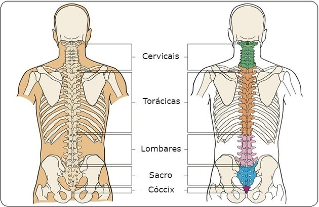
The spine is formed by vertebrae that are linked together by joints, which makes the spine very flexible. It has curvatures that help to balance the body and cushion shocks during movements.It consists of 24 independent vertebrae and 9 that are fused. See in the table below how they are grouped:
| Vertebrae | Characteristics |
|---|---|
| Cervicals | There are 7 neck vertebrae, the first (atlas) and the second (axes) favoring the movements of the skull. |
| Thoracic or dorsal | There are 12 and articulate with the ribs. |
| Lumbar | These 5 vertebrae are the largest and the ones that support the most weight. |
| Sacrum | These 5 vertebrae are called sacral, are separated at birth and fuse later to form a single bone. It is an important support point for the pelvic girdle. |
| Coccyx | There are 4 small coccygeal vertebrae that, like the sacral vertebrae, become united in a single bone in early adulthood. |
Chest
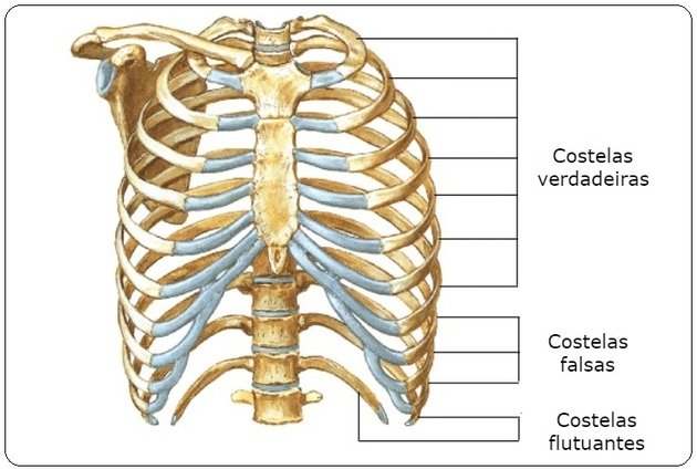
The chest consists of 12 pairs of ribs connected to each other by the intercostal muscles. They are flat and curved bones that move during breathing. The ribs are connected to the thoracic vertebrae at the back.
Previously, the first seven pairs of ribs (called true) are attached to the sternum, the next three (false) are attached to each other, and the last two (floating) pairs are not attached to any bone. The sternum is a flat bone that attaches to the ribs through cartilage.
Hyoid bone
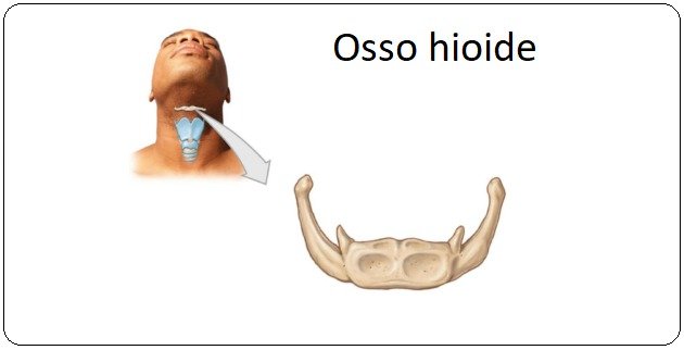
The hyoid bone is U-shaped and acts as a support point for the tongue and neck muscles.
Appendicular Skeleton
The appendicular skeleton includes the “appendages” of the body. They correspond to the bones of the upper and lower limbs.In addition, the appendicular skeleton has the bones that connect them to the axial skeleton, the so-called scapular and pelvic girdles, in addition to ligaments , joints and joints.
Shoulder girdle
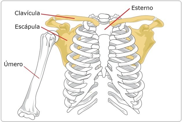
The scapular waist is formed by the clavicles and scapulas.
The clavicle is long and narrow, articulating with the sternum and at the other end with the scapula, which is a flat, triangular bone articulated with the humerus (shoulder joint).
Upper limbs
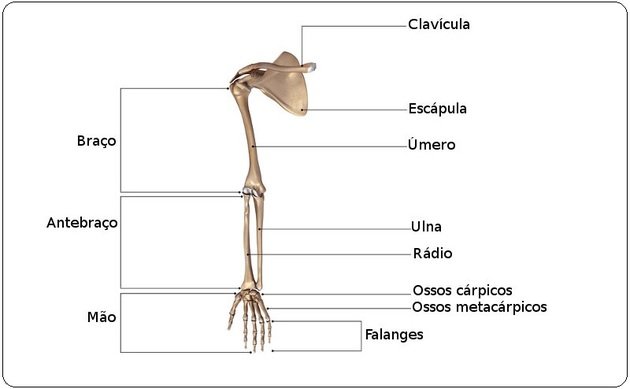
The upper limbs correspond to the arms, where there is the humerus, which is the longest bone in the arm. It articulates with the radius, which is the shortest and lateral, and also with the ulna, flat and very thin bone.The bones of the hand are 27, divided into carpus (8), metacarpal (5) and phalanges (14).
Pelvic Girdle
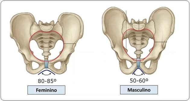
The pelvic girdle is formed by the hip bones, the iliac bones (consisting of the fused ilium, ischium and pubis) and are firmly attached to the sacrum.The union of the iliac bones, the sacrum and the coccyx form the pelvis, which in women is wider, less deep and with a larger cavity. It is this formation that allows the pelvis to open at the time of delivery for the baby to pass.
Lower members
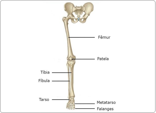
The bones of the lower limbs are responsible for supporting the body and movement. For that, they have to support the weight and maintain balance.
See the table below for the characteristics of the lower limb bones:
| Bones of the lower limb | Characteristics |
|---|---|
| Femur | It is the longest bone in the body. It has a rounded head to fit the pelvis. |
| Patella | It is a sesamoid bone, articulated with the femur. |
| Tibia | It supports almost all the weight in the lower part of the body. |
| Fibula | It is a weaker bone, connected with the tibia helps to move the foot. |
| Bones of the foot | The feet have 26 bones divided into: tarsi (7), metatarsals (5) and phalanges (14). |
Ossification and Bone Remodeling
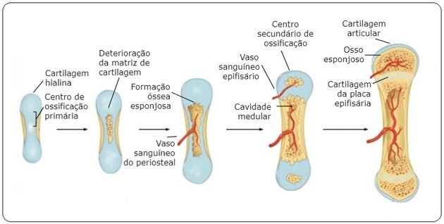
The bone formation process starts around the first 6 weeks of life and ends at the beginning of adulthood. However, the bone continually undergoes a remodeling process, where part of the existing tissue is reabsorbed and new tissue is formed.