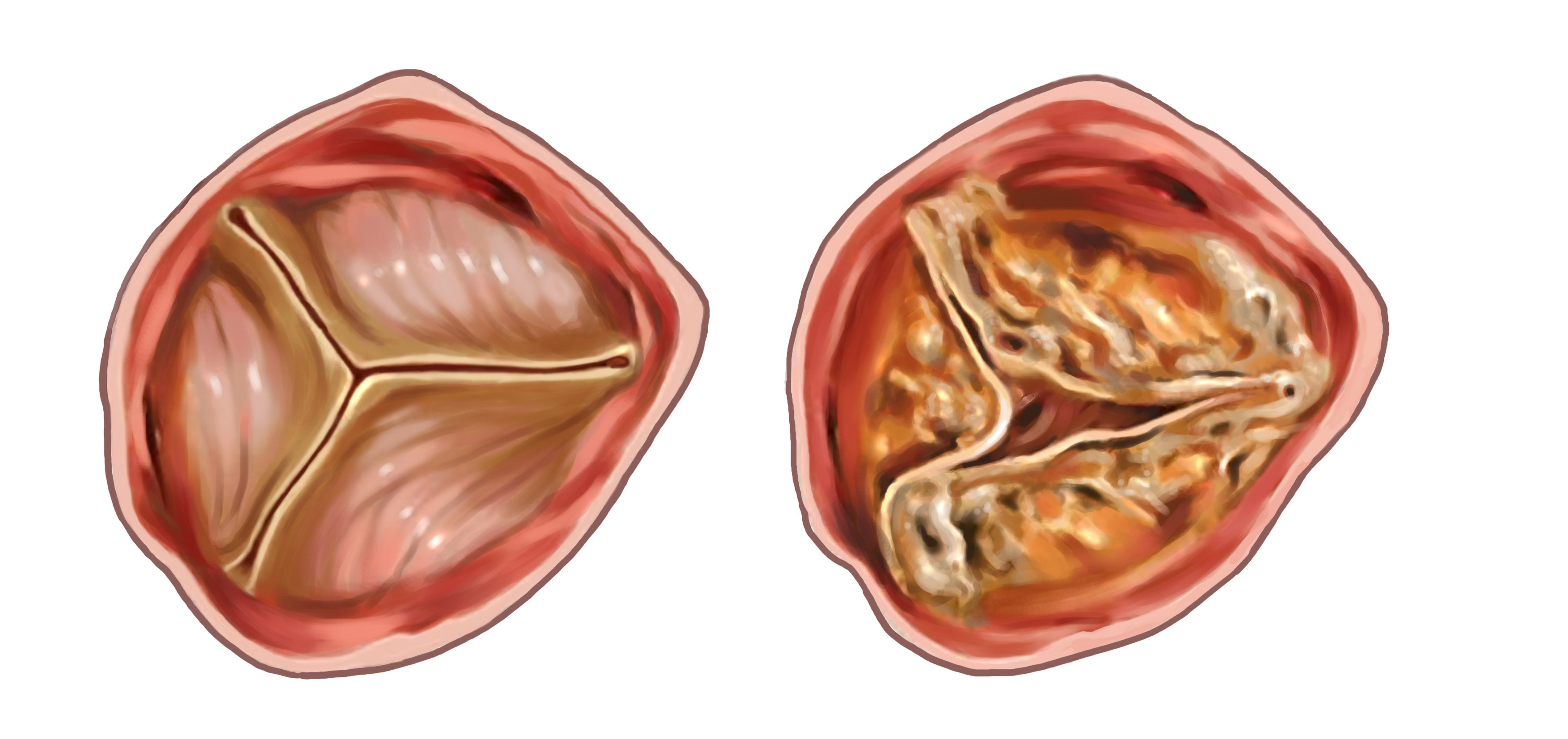
Tricuspid stenosis is rare as an isolated lesion. Almost always it accompanies rheumatic involvement of other cardiac valves, and it is of clinical significance in only about 5 per cent of patients with rheumatic heart disease. Seventy to 80 per cent of patients with tricuspid stenosis are female, and most often they have accompanying disease of both the mitral and aortic valves.
Rare causes of tricuspid stenosis include carcinoid heart disease. endomyocurdial fibroe/astosis, congenital malfor- mations, and obstruction from a right atrial myxoma. The primary abnormality in tricuspid stenosis, like that in mitral stenosis, is mechanical obstruction to atrial emptying.
This results in an elevated right atrial pressure and a pressure gradient across the tricuspid valve during diastole. It is important to measure simultaneous pressures in the right atrium and right ventricle at cardiac catheterization, because signs of peripheral venous congestion may occur with substantially lower pressure gradients (less than 10 mm Hg; often only 3 or 4 mm Hg) than are observed with mitral stenosis. Generally, the cardiac index is reduced substantially.
Tricuspid stenosis should be suspected particularly in the patient who has signs of multivalve disease and severe right-sided venous congestion but no evidenced marked pulmonary hypertension. The patient may also be relatively free of signs and symptoms of pulmonary venous congestion, despite the presence of mitral valve stenosis. This feature may be due to a limiting effect of this lesion on the cardiac output (a protective effect of the tricuspid stenosis) which may prevent sudden augmentation of blood flow into the pulmonary vascular bed. Such an effect would help explain the unusually pro- longed clinical course seen in some patients with tricuspid stenosis. On physical examination, the mean jugular venous pressure is elevated, and in patients with sinus rhythm there is a very sharp, prominent A wave which reflects right atrial contraction against the stenotic valve. There is a characteristic slow fall in the jugular V wave (delayed Y descent), caused by the obstruction to right.
On palpation of the precordium, the right ventricle is not enlarged, and a diastolic thrill may be palpable at the lower left sternal border. The diastolic murmur of tricuspid stenosis will often be missed unless specifically searched for at that location. It resembles that of mitral stenosis, having a low-pitched rumbling quality often beginning with a tricuspid valve opening snap (some- what later than a mitral opening snap), and an early diastolic decrescendo murmur is followed by presystolic accentuation if sinus rhythm is present. A characteristic feature in differentiating the murmur from that of mitral stenosis is augmentation in the intensity of the tricuspid rumble during inspiration, or during the Miiller maneuver (attempted inspiration against a closed glottis).
This augmentation is due to the transitory increase in right atrial filling and tricuspid valve flow, which results from the negative intrathoracic pressure. Both these maneuvers diminish the murmur of mitral stenosis. There may be an associated abnormal increase in the venous pressure during such inspiration in tricuspid stenosis. When atrial fibrillation is present, the murmur is even more difhcult to detect, because it may occur only in early diastole and can be confused with the murmur of pulmonic regurgitation.
The electrocardiogram, if sinus rhythm is present, exhibits peaked P waves in leads II and VI, evidence of right atrial enlargement, but atrial fibrillation is often present. There is no evidence of right ventricular hyper- trophy. On the chest roentgenogram, in addition to the findings caused by associated valvular lesions, there is enlargement of the right atrium.
Surgical treatment is not indicated for mild tricuspid stenosis, but if it is severe or if the patient must undergo operation for a mitral valve lesion, surgical treatment may be undertaken. The tricuspid valve is usually not favorable f’or valvuloplasty, and most often a tricuspid valve prosthesis is inserted. Sometimes the tricuspid stenosis is not detected prior to a mitral valve operation, and is recognized only when unexpected signs of right- sided congestion develop postoperatively.
