Rift Valley fever is a toxic generalized febrile illness of short duration, accompanied by headache, photophobia, myalgia, anorexia, prostration, and leukopenia. It is caused by a mosquito-borne pantropic virus that is epizootic in domestic animals and enzootic in wild game, restricted, as its name implies, to East and South Africa.
Etiology.
Rift Valley fever virus -ranks’ second only to yellow fever virus in historic order of isolation as a filterable arthropod-borne cause of disease. Although first isolated from blood and tissues of diseased sheep and cattle in 1930 in East Africa, it was not isolated from wild mosquitoes until 1944. In propagation in and experimental transmission by mosquitoes, Rift Valley fever virus is species-selective.
Like the other arboviruses, Rift Valley fever virus is small, in the 23 to 50 m/x range. It can be preserved indefinitely in the lyophilized state. Appropriately prepared suspensions of infected material contain complement-fixing and hemagglutinating antigens. Very high titer antigens can be prepared by extraction of infected brain material by organic solvents.
Human infection results not only from mosquito transmission but from contact with tissues and secretions of sick animals and especially from handling the virus in the laboratory. Because of its high infectivity for man and widespread endemic and epidemic involvement of domestic and wild animal populations of Africa, to which its apparent natural range is limited, it is one of the few viruses prohibited in the United States, even for experimental work.
Epidemiology of Rift Valley Fever.
The investigations that led to the isolation of Rift Valley fever virus in 1930 as a cause of disease in domestic animals and associated human beings in East Africa are classic in veterinary medicine. By moving afflicted fiocks of sheep from mosquito-infested environments to higher elevations where mosquitoes were few, and by experimentally holding susceptible sheep under mosquito nets in perfect health while adjacent freely exposed pasture animals fell victims to the infection, indirect evidence accumulated that implicated mosquitoes as Rift Valley fever virus vectors.
The variety of species and their distribution indicate that the virus is enzootic in wild game and other animals that abound in the zone of incidence. Following unusually heavy rains, which promote excessive mosquito production, the virus spills over from its wild reservoir into domestic herds and flocks, and the associated mosquito transmission results in epizootics that have been recorded many times in East and South Africa.
. The first outbreak of Rift Valley fever recognized in South Africa occurred in 1951. Several have occurred since that time. Although livestock was affected primarily, associated cases resulted in persons who handled sick animals or their flesh. Antibody surveys indicate that virus activity is widely dispersed in East and South Africa, where natural human exposure seems to be uncommon.
Histopathology.
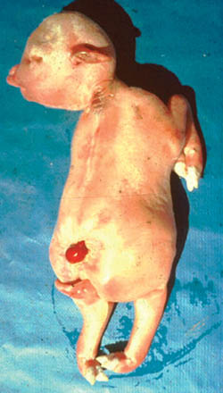
The characteristic macroscopic and microscopic pathologic changes of this disease in sheep led veterinarians to label it initially as enzootic hepatitis. It was its nonfatal occurrence in man, in whom the pathologic processes are obscure because of the paucity of postmortem tissue, that caused adoption of the geographic term.
The pathognomonic lesion is necrosis in the liver, which in lambs, calves, and inoculated mice may be so extensive that even lobular architecture is lost. The process involves focal hyaline degeneration of the cell cytoplasm that ultimately results in development of cytoplasmic inclusion. Nuclei of hepatic cells are also attacked, and acidophilic intranuclear inclusions appear. As necrosis proceeds, there is massive accumulation of polymorphonuclear leukocytes around cell detritus, and erythrocytes may pack the sinusoids Toxic degeneration, such as sloughing of the tubular epithelium in the kidneys and erythrocytic extravasation in the viscera, even with hemorrhagic enteritis, may occur. The process in Rift Valley fever in sheep and :arde is very similar to that seen in yellow fever and Kyasanur Forest disease in man. the tm latter being respectively mosquito-borne and tick-borne group B arbovirus infections in man.
You Must Understand Rift Valley Fever Virus Clinical Manifestations.
Whatever the mode of virus trams mission may be, it appears that some degree of overt disease occurs in most human infections.The onset may be gradual over a number of hours, but it is usually sudden with high fever, photophobia, and intractable headache after an incubation period ranging from three to six days. In severe disease there are extreme prostration and generalized shifting myalgia, which may be most painful in the back and extremities. Anorexia is common. Pain occurs in the epigastrium. Nausea and vomiting may also occur.
The temperature ranges from 101 to 104° F. and often presents a “saddle back” curve in the course of acute illness, which lasts from two to six days, averaging something more than three days. Bradycardia relative to the level of fever is commonly observed as it is in a number of other pantropic toxic arbovirus infections, such as yellow fever. Leukopenia in the range of 4000 cells per cubic millimeter is characteristic and parallels the duration of the fever. It should be remembered that malaria parasites have appeared during the febrile phase in patients with Rift Valley fever. There is no response to anti- malarial drugs, and the fever does not subside until shortly after viremia ceases.
Convalescence from Rift Valley fever usually progresses rapidly to complete recovery, although severe prostration during the acute illness may require prolonged convalescence of ten days or more. Vascular changes and hemorrhage may also result from infection with this virus.
Complications.
The complication most frequently reported in man is a central serous retinopathy with associated central scotoma. Examination of the fundus shows exudative areas of variable size involving the macula- that may result from thrombosis of the vessels. These exudative areas may last for several weeks, changing from a white mass to crenated shrinkage and ultimate disappearance. Although most patients recover completely and have a return of normal vision, retinal detachment has occurred. This is the most serious sequel to Rift Valley fever. In Africa any sudden visual defect following an acute febrile illness should suggest the possibility pf Rift Valley fever complicated by serous retinopathy.
Diagnosis.
Occupational or recreational exposure in potentially infected areas of Africa, association with diseased animals or their tissues, and laboratory exposure to the virus are the only leads to a tentative clinical diagnosis in a patient with an acute fever, myalgia, and leukopenia. Isolation of the virus by intracerebral inoculation of suckling or weanling mice with blood collected during the first three days of illness is the most certain way to establish a diagnosis. Rift Valley fever virus has a short incubation period, so the mice should sicken within three days after inoculation. Identification, using immune reference serum, can be accomplished by complement- fixation. hemagglutination-inhibition, or neutralization tests.
Prevention And Treatment of Rift Valley Fever
The original animal experiments that implicated mosquitoes as the vector demonstrate the value of elimination of exposure to mosquito bite in potentially infected areas by use of mosquito nets, screening, and repellents. This should be kept in mind by anyone in pursuit of game animals in East and South Africa.
Serial intracerebral passage of one strain of Rift Valley fever virus in mice has increased neurotropism at the expense of hepatotropism. This attenuated strain is now effectively used to immunize sheep, cattle, and other susceptible animals. No evaluation of the vaccine has been reported in man. A formalin-killed virus vaccine for human prophylaxis is presently under study.