Q fever is a self-limited rickettsial infection characterized by fever, headache, and constitutional symptoms, associated, in approximately half the patients, with a pneumonitis. It is unique among the rickettsial diseases of man in that human infection is most commonly acquired by inhalation of the agent rather than by contact with an arthropod vector.
Etiology.
The disease is caused by Coxiella burnetii, a rickettsial agent having the general biologic characteristics of this class of microorganisms but possessing, in addition, a resistance to desiccation and to exposure in dusts and soils that is unique among the rickettsiae. The organism may be propagated in embryonated eggs and in mice, hamsters, and guinea pigs, and has frequently infected laboratory personnel engaged in isolation studies on clinical material. Both patients and animals develop agglutinating and complement-fixing antibodies for the agent. On the other hand, C. burnetii, unlike most of the other rickettsiae, does not stimulate the production of Weil-Felix Proteus agglutinins in man.
Incidence and Distribution.
The true incidence of the disease in the human population is impossible to determine because the majority of infections are undiagnosed. Isolated serologic surveys have revealed that many persons exposed to infection in sheep and cattle ranches, abattoirs, meat packing plants, wool processing plants, etc., present serologic evidence of past infection. More detailed investigation of animals and arthropods on a worldwide basis has shown C. burnetii to be ubiquitous in distribution except for the countries of Denmark, Finland, Ireland, the Netherlands, Norway, and Sweden. In the United States the disease, first recognized in the states of Montana and California, is now recognized as prevalent in most of the states in which sheep and cattle are produced. Small numbers of cases have been reported for most of the remaining states.
Epidemiology.
The epidemiology of the disease is complex because it involves two major patterns of transmission. The first pattern, described in Australia, is a disease cycle in wild animals with transmission of the agent from animal to animal by a tick vector. Although the species of animal and arthropod vary from country to country, such a cycle has been demonstrated in Australia in two forms, bandicoot-tick-cattle and kangaroo- tiek-sheep. In both these cycles the agent can be transmitted indefinitely as an inapparent infection in the wild reservoir (bandicoot or kangaroo) by ticks, but it may also be transmitted laterally by arthropod to a domestic animal in close contact with man. In these and similar cycles recognized in other parts of the world, C. burnetii, like the other rickettsial agents of human disease, is vector- transmitted.
However, Q fever patients rarely give a history of tick bite. Human infection with the agent has now been shown to occur almost exclusively by a second transmission pattern capable of sustaining itself independently of the wild animal cycle. The reservoirs of infection in the second pattern are animals domesticated by man, principally cattle, sheep, and goats, in which C. burnetii produces only an inapparent or mild infection. In sheep, Q fever organisms are excreted in very large numbers in placental tissue, and to a lesser degree in birth fluids, milk, and feces. In the cow, and probably the goat, excretion occurs mainly through the placenta and milk. With all infected animals, the period of parturition is associated with the formation of a primary infectious aerosol, easily demonstrated by air-sampling studies.
Such aerosols infect other cattle in the herd and also the human population in direct contact with the animals. Further, because contaminated clothing, wool, hides, bedding, and soil may be the source of secondary aerosols, the infections may be transmitted via these vehicles at considerable distances from the infected cattle; in certain circumstances these distances are’ measurable in miles.
The unique resistance of C. burnetii to prolonged exposure in nature contributes to the spread of the agent by such infectious microenvironments. Within California, where the local epidemiology of the disease has been studied in great detail, sheep are the major reservoir for human infection in the northern part of the state and cattle in the southern part. In the latter area milk may be a vehicle of infection if consumed raw. Although the pulmonary route is the most important portal of access of the agent to man, the transmission of the rickettsia from man to man by this method ‘is rare, despite the occurrence of an infectious pneumonitis in some patients.
Because the mortality rate is low, postmortem studies have been limited. In those patients having a pneumonitis, the histopathology is similar to that seen in the viral pneumonias and psittacosis. Recent studies in patients have directed attention to hepatic pathology during the acute phase of the disease, demonstrable both in biochemical abnormalities of liver function (elevated cephalin-cholesterol flocculation, alkaline phosphatase, and thymol turbidity tests) and in histologic abnormalities in biopsy specimens (focal inflammation and granulomas). Q fever endocarditis, producing valvular vegetations from which rickettsiae may be isolated, has also been described.
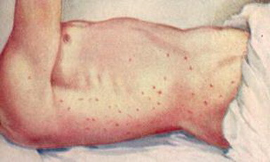
After an incubation period of 9 to 20 days following respiratory exposure, most patients complain of an abrupt onset of fever, headache, muscle pains, and severe malaise. The temperature may rise as high as 104° F. and remain elevated, with considerable fluctuation, for one to three weeks. Occasionally, patients may suffer a prolonged fever of several months’ duration. In contrast to the other rickettsial infections, there is no rash. In approximately half the patients there is roentgenographic evidence of pneumonitis, manifested clinically as a slight, nonproductive cough developing in the second week of fever.
Diagnosis.
Q fever should always be suspected in a patient having a febrile illness for which no obvious cause can be found. If the patient’s occupation brings him into contact with sheep, cattle, or goats, or byproducts such as wool or hides, particular care should be exercised to exclude Q fever from consideration. The presence of Q fever should be suspected in any patient in whom the differential diagnosis includes viral pneumonia, psittacosis, primary atypical pneumonia, pul- nionary mycotic disease, or comparable infections. Furthermore, recent data would also support consideration of Q fever in the differential diagnoses of endocarditis and of hepatitis with or without jaundice.
Diagnostic laboratory studies usually must be limited to serologic studies because of the extensive history of accidentally acquired laboratory infections resulting from attempts at isolation. Either complement-fixation or agglutination tests may be employed and with either test a fourfold or greater rise in titer of antibody may be expected to occur between the first and fourth weeks of illness. Recent reports have emphasized the importance of both strain and phase variation in the selection of antigens for diagnostic use.
Treatment and Prognosis
The tetracyclines or chloramphenicol are both effective in treatment of lower still in those treated with antimicrobial drugs. Therapy should be continued for approximately one week even though the patient usually becomes afebrile within 48 hours. Patients occasionally may experience relapses after treatment, and when this complication occurs additional drug therapy should be administered. Prognosis is less favorable for the rare patient in whom Q fever endocarditis develops. In some of these patients the disease has been reported to have been unresponsive to antimicrobial therapy.
Prevention
Experimental lots of yolk-sac vaccines have been effective in prevention of clinical disease in volunteers experimentally infected via the respiratory route. Because this vaccine is not commercially available, control measures are limited to minimizing exposure to the agent. In particular, milk from cattle, sheep, and goats in endemic areas should be pasteurized or boiled before use. Because rickettsiae are excreted in the sputum and urine of patients, these materials should be disinfected^ by autoclaving to prevent secondary infection in hospitals.
