Varicella zoster virus are now considered the consequence of the activity of a single virus and the dissimilarity of the clinical features of the two syndromes as a reflection of differences in the response of the human host to the same agent. Although the mechanism of pathogenesis is incompletely understood, varicella may constitute the response of the nonim-mune host and herpes zoster that of the partially immune person.
Definition of varicella.
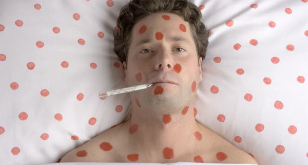
Varicella is a highly contagious disease characterized by a generalized vesicular exanthem developing in crops over a period of a few days. Usually benign in children, in adults it may be accompanied by severe symptoms.
Herpes zoster is an infectious process associated with the appearance of a circumscribed vesicular eruption of the skin or mucous membranes. The localized eruption, often involving one or more dermatomes, reflects a concurrent inflammatory process in related dorsal root ganglia or extramedullary cranial nerve ganglia.
Although varicella and herpes zoster are usually considered as discrete syndromes, the patient with zoster not infrequently shows evidence of generalization of the cutaneous process, and rarely a zosteriform concentration of vesicular lesions occurs in the patient with varicella.
Etiology.
The virus is highly host-specific and has been transmitted experimentally only to man. Electron microscopic examination of varicella or zoster vesicle fluid reveals the virus particles as round bodies 210 mju in diameter. Isolation of virus from vesicular lesions of cases of varicella and zoster was accomplished by Weller < 1953) in cultures of human tissues. Employing viruses thus grown in vitro, the similarity of agents recovered from the two clinical syndromes was established. Now commonly termed varicella-zoster virus or V-Z virus, the agent is classified in the herpes virus group, and contains nucleic acid of the deoxyribose type. Varicella-zoster virus is distinct from, and not to be confused with, the atidougfi possessing a similar ultrastructure, and having minor antigens in common. In tissue culture the virus produces a cytopathic effect comparable to that seen in vivo with the development of multinucleate giant cells and eosinophilic intranuclear inclusions.
Incidence and Epidemiology.
Varicella occurs at any age. In temperate regions, the greatest prevalence is observed between the ages of two and eight years. Fortunately, relatively few cases occur in the adult population, which is generally immune. However, in some tropical areas varicella is primarily a disease of adults. Varicella in temperate areas commonly presents in epidemic form during the cooler months, whereas zoster is a sporadic disease occurring throughout the year. Although cases of zoster occasionally are seen in infants and children, zoster is charae-teristicaliy a disease of adults, attack rates increasing with age. In England, a recent study indicates tha: approximately half of the people achieving age 85 will have experienced an attack of zoster (Hope-Simpson, 1965).
Varicella is the consequence of contact with a pre-existing case gy, as by von Bokay (1909). of contact with a person with herpes zoster. Zoster, on the other hand, often appears in the absence of contact with an external source of virus, and is believed to be the result of reactivation of virus lying latent in the body. Careful investigation of zoster occurring in children yields a history of prior varicella in a high percentage, evidence consistent with the concept of viral latency. Occasional patients with zoster give a history of recent contact with an external source of virus; such contact is probably coincidental, for epidemiologic studies fail to reveal an increased prevalence of zoster during periods when varicella is epidemic.
Pathology and Pathogenesis.
In varicella the initial site of viral replication is not known, but may be in the respiratory tract. Thereafter, viremia probably follows with the initiation of focal lesions that apparently enlarge by spread of virus from cell to contiguous cell. As described by Tyzzer (1906), the initial changes in the skin take place in the endothelium of capillaries in the corium. Cells in the basal and prickle layers undergo ballooning degeneration, and fluid accumulates, the intact stratum corneum forming the roof of the vesicle. Nuclear changes with margination of chromatin and the appearance of intranuclear inclusions are characteristic of the reaction of infected cells. In the skin, multinucleate giant cells appear, each nucleus containing an inclusion. Virus is present in large amounts in the clear fluid of the young vesicles; as the vesicle fluid becomes cloudy with accumulating inflammatory cells and debris, the viral content declines. In fatal cases, focal areas of necrosis with associated specific nuclear changes occur in many organs.
In zoster, the cutaneous lesion morphologically is like that of varicella. The posterior root ganglion corresponding to the cutaneous site is acutely involved, and intranuclear inclusions have been demonstrated in ganglion and in satellite cells. Extension of the inflammatory process to the pos-terior horns and less often to the anterior horns of the cord may occur; virus has been recovered from the cerebrospinal fluid, and a pleocytosis in-the fluid is not unusual.
Partial degeneration of the dermal nerve network occurs in the affected dermatome, and there may be loss of functional integrity of the sensory afferent nerves. Zoster most commonly involves areas of skin innervated by the thoracic ganglia (50 per cent), the cervical ganglia (15 per cent), and the ophthalmic branch of the gasserian ganglia (10 per cent). The pathogenesis of zoster is not understood; in the “idiopathic” type, no reason for activation is apparent. In other instances, a trigger mechanism in the form of trauma, injections of drugs such as arsenicals, or concomitant disease such as tuberculosis or malignancy (particularly lymphoma and chronic leukemia), may be evident. Zoster is a relatively common iatrogenic complication in the recipient of immunosuppressive therapy.
They are numerous on the trunk and face and relatively sparse over the extremities; mucosal surfaces may also be involved. The typical lesion develops within a few hours from a small macule to a fragile, slightly elevated, dewdroplike vesicle with a red areola’. With involution, it becomes crusted, and frequently is secondarily infected. The vesicle is delicate and easily broken but is not unilocular as is sometimes stated. Pruritus may be severe. The duration and degree of fever parallel the severity of the eruptive process. Lesions are usually more numerous in regions of per-existing inflammation.
Because of the appearance of new crops, the presence of lesions in various stages of development in a single area is characteristic. Those appearing in the final crop may not progress beyond the maculopapular stage.
In infants, in children with leukemia or on steroid therapy, and in adults, varicella may be serious and may have a fatal outcome. The rash may persist, may become more extensive and hemorrhagic, and a specific pneumonitis demonstrable roentgenographically as a bilateral nodular infiltration may develop. There may be a focal hepatitis. Varicella pneumonia occurs primarily in adults. After recovery, serial roent-genographic examinations may reveal the development of small nodular calcifications scattered throughout the lung fields. Postvaricella encephalitis is a rare and grave complication of mild as well as of severe cases. In a different category is the disturbing but transient cerebellar type of ataxia occasionally seen in children after varicella.
Defination of Herpes Zoster.
Pain and paresthesia often precede the appearance of skin lesions by a few days. The individual lesions evolve as in varicella, singly or irregularly in almost confluent patches on an erythematous base, but are concentrated over the area of distribution of one or more spinal nerves or the sensory division of a cranial nerve; thus, a unilateral bandlike eruption is characteristic. However, in one third of cases scattered cutaneous lesions may appear on other areas of the body. (See also Disorders of the Nervous System and Behavior.) The regional nodes enlarge; rarely this enlargement may precede the pain and skin involvement by a few days. Systemic complaints in the form of malaise, headache, and fever are present in a small percentage of cases.
In the majority of patients with zoster, spontaneous resolution of the cutaneous lesions and disappearance of pain occur within two weeks. However, in a minority, postherpetic neuralgia persists as a distressing and refractory consequence of infection. Scarring of the cornea may result from ophthalmic zoster.
Varicella Zoster Virus Diagnosis.
Differentiation of varicella from mild smallpox on clinical grounds may be difficult; the demonstration of the characteristic multinucleate giant cells with intranuclear inclusions in stained scrapings of young vesicles or biopsy materials will establish the presence of either varicella-zoster or herpes simplex virus and will eliminate variola or vaccinia virus from consideration. Other entities with a generalized vesicular eruption to be considered in differential diagnosis include eczema vaccinatum and eczema herpeticum, rickettsialpox, and certain Coxsackie virus infections. An atypical zoster eruption may be clinically indistinguishable from the lesions of recurrent herpes simplex.
Pain in the pre-eruptive stage of zoster may suggest a variety of conditions such as pleurisy or appendicitis. To establish a specific diagnosis, virus may be isolated by the inoculation of appropriate cell cultures with fluid from newly appearing vesicles; cutaneous lesions that are purulent or crusted are usually virolo-gically negative. The demonstration of a rising titer of complement-fixing antibodies for varicella-zoster antigen is helpful, but must be interpreted with some caution owing to antigenic components shared with the herpes simplex viruses.
Varicella Zoster Virus Treatment.
There is no specific treatment for varicella or zoster. Therapy is symptomatic and is directed at relief of local discomfort and the control of secondary infection. Prolonged mechanical ventilation may be required to maintain oxygenation of the patient with varicella pneumonia. It is to be noted that critical evaluation of therapeutic approaches to the treatment of zoster is difficult because of the self-limited and variable nature of the acute process. Postherpetic pain may ultimately require neurosurgery.
Prevention.
No vaccine exists. Ordinary human immune serum globulin administered to children exposed to varicella does not prevent infection, but may modify the severity of the illness. However, recently it has been established that immune globulin prepared from selected donors recovering from herpes zoster will prevent infection if administered to susceptible children within 72 hours of exposure.