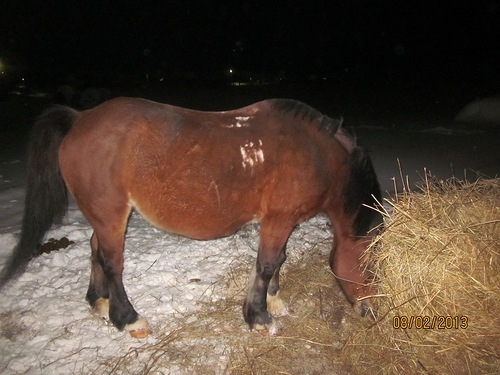
Glanders, or farcy, is an infectious disease of horses, mules, and donkeys caused by Malleomyces mallei. The infection is occasionally transmitted to man, and is characterized by an acute fulminant febrile illness or a chronic indolent disease with abscesses of the respiratory tract or skin. Farcy refers to the nodular abscesses observed in skin, lymphatics, and subcutaneous tissues.
Etiology.
Glanders was described by Aristotle about 330 B.C., and the occurrence of the disease in horses was observed by Apertos about 375 a.d.Royer’s (1837) monograph on glanders in man remains the classic description of the disease. The micro-organism responsible for glanders was isolated in 1882. It is a gram-negative bacillus cultured aerobically on ordinary nutrient media. It is variously called Allomyces mallei, Bacillus mallei, or Pfeiferella mallei. M. mallei is nonmotile. It elaborates a specific antigen (mallein) upon lysis that is used as a skin test material for diagnostic purposes. The bacillus is antigenically separable from M. pseudomallei, which causes melioidosis. When grown on potato slices or potato infusion agar, M. mallei produces a brown pigment resembling that of Pseudomonas aeruginosa. The bacillus produces fatal infection experimentally in guinea pigs and hamsters but will not cause disease in rats, cattle, hogs, or fowl.
Incidence and Prevalence.
Glanders has probably never been a common disease in man. It occurs almost exclusively in persons handling horses and, therefore, is an occupational disease. It has been reported from all parts of the world. It has been eradicated in the United States and the United Kingdom, but may still occur in Asia and South America.
Epidemiology.
Glanders is a communicable disease among horses, and it may occur sporadically in other animal species in contact with horses. In horses, glanders is manifested by nasal symptoms and abscesses and by cutaneous nodules or abscesses (farcy). Glanders is transmitted to man by direct contact with infected horses, the bacillus preferentially invading areas of abraded or injured skin. Experimental infection can be induced in animals by inhalation of the bacillus, and certain laboratory-acquired infections indicate that infection may develop in man following inhalation of the micro-organism. Unlike melioidosis, glanders can be transmitted from person to person.
Pathology and Pathogenesis.
Characteristically glanders is associated with cellulitis, necrosis, abscess, and thromboses with septic embolization. Healing occurs by fibrosis, and, rarely, by calcification.
Clinical Manifestations of Glanders The Americans.
Glanders may occur as a fulminant acute febrile disease, as a chronic indolent and relapsing disease, or as a subclinical occult infection detectable incidentally at postmortem examination or by serologic test. The two principal features of glanders are (1) nasal cellulitis and necrosis-producing septal perforation, palatal and pharyngeal ulceration; or (2) cutaneous cellulitis, vesiculation, and ulceration at the site of inoculation of the bacillus into the skin, followed by lymphangitis with nodular abscesses along the lymphatics and lymphadenopathy (farcy).
Pulmonary involvement is common in glanders, producing pneumonia, abscesses, pleural effusion, and empyema. Occasionally, in chronic indolent glanders, small nodular granulomatous lesions may be found in the lungs. Hilar lymph node enlargement is common in pulmonary glanders.
When glanders becomes disseminated, destructive polyarthritis, subcutaneous and muscular abscesses, osteomyelitis, meningitis, and pustular skin lesions, particularly over the joints and face, are seen. Prostration and stupor may develop, and are followed by death in two to three weeks.Chronic glanders may be punctuated by recurrent acute relapses with bacteremia, often resulting in a fulminant course and death. Amyloidosis may be a complication of chronic glanders.Fever and chills, headache, and backache are common in acute glanders. Leukopenia or a normal leukocyte count is the rule, but leukocytosis has been observed.
Diagnosis.
There are no pathognomonic clinical features of glanders. The nodules along lymphatics resemble those seen in sporotrichosis. The respiratory lesions may be difficult to distinguish from other ulcerative infections of the nose and mouth. The cutaneous lesion often resembles streptococcal cellulitis. The multiple abscesses mimic many mycotic and staphylococcal infections and are difficult to distinguish from those of melioidosis. The acute fulminant illness may resemble typhoid fever or disseminated tuberculosis. The diagnosis of glanders can be established by combination of a history of exposure to horses, isolation of M. mallei, serologic tests (agglutination, complement-fixation), and skin test with sterile culture filtrate (mallein). Inoculation of infected material into guinea pigs may facilitate identification of the micro-organism.
Treatment of Glanders In Horses.
There are reports of successful treatment of glanders with sulfonamides. Tetracycline, chloramphenicol, and streptomycin may be useful, but there is little clinical experience with these drugs in glanders. Because of the serious prognosis and the lack of documented experience with drug therapy, it is advisable to administer daily streptomycin (1.0 gram) in association with a sulfonamide or a tetracycline until all evidences of disease have disappeared. Incision, drainage, and excision of abscesses must be done with caution, for the infection may be disseminated by such manipulation.
Prognosis.
Although a few patients have been cured with chemotherapy, the effect of treatment on the mortality rate is not known. More than 90 per cent of patients with glanders will die from the disease if untreated. However, the frequency of occurrence of occult or subclinical glanders is not known.
Prevention.
Infected horses have been identified by skin tests with mallein and by serologic tests. Destruction of infected animals has eliminated glanders as a public health problem in the United States and most other countries.
