Dichroism, a property possessed by some doubly refracting crystalline substances of appearing of different colours, when viewed in polarised light, the difference of colour depending upon the direction in which the luminous vibrations take place.

The Dichroism, of crystals can be observed or tested by means of a simple instrument called a dichroscope or `dichroo-scope’. It is simply a cleavage rhombohedron of Iceland spar with a weak magnifying lens. The crystal is held in a good light opposite one end of the instrument and on looking through it 2 images of the square hole are seen just touching each other.
If the crystal is dichroic these will be of different colours and the colours will change if the dichroscope is rotated between the fingers. The most remarkable dichroic substances are the magnesian mica from Vesuvius, the tour-maline and ripidolite, also the sapphire and ruby.
The phenomena of dichroism are best seen in crystals with 2 axes of double refraction, notably in which crystallises in 6- or 12-sided prisms; these prisms, when seen along the axis, are of a deep blue colour, but when viewed in a direction perpendicular to it assume a yellowish-brown colour. Tourmaline is another notable example, being blood-red when viewed along the axis, but yellowish-green when viewed at right angles to it.
The Function of Circular Dichroism
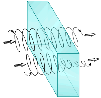
The circular dichroism (CD), measured as a function of the wavelength, is the difference in absorption between the right component (R-CLP) and the left component (L-CLP) of the circularly polarized light.
This difference can be detected when a molecule contains one or more chiral groups that absorb light – the so-called chiral chromophores.
Circular dichroism (CD) = ΔA (λ) = A (λ) L-CPL – A (λ) R-CPL , where λ is the wavelength.
When chiral chromophoric components are present, one of the circularly polarized light states is absorbed with greater intensity than the other. At the same wavelength, a CD signal can therefore be positive or negative, depending on whether the left L-CLP component is absorbed more or less than the R-CLP right component (positive or negative signal).
The CD Chirascan spectrometers measure the absorption of the R-CLP right component and the L-CLP left-hand component alternately at each wavelength to calculate the difference or the CD signal. Therefore, the CD spectra vary according to the different absorptions, leading to the visualization of different peaks, positive when L-CLP> R-CLP or negative when L-CLP <R-CLP, depending on the different wavelengths.