Different Types of Heart Failure In Human Being are being discussed in this article.The heart is part of an elaborate cardiopulmonary system that is designed to satisfy the metabolic needs of the tissues with respect to oxygen, substrates, and nutrients not only at rest but also during activity. This system includes a sophisticated regulatory apparatus for apportioning the cardiac output according to the physiologic priorities of the moment, e.g., muscular exercise, heat loss, digestion. The same regulatory mechanisms that satisfy physiologic priorities in health also serve to protect vital organs, e.g., the heart and brain, when cardiac output is seriously compromised.

According to its structure and function, the heart may be examined as a pump, as a muscle, or as a component of the cardiopulmonary system. And when one of the two ventricles fails, the consequences may be viewed in the same way. Because of its central role in meeting the needs of the body, and because the pulmonary circulation is lodged between the two ventricles, heart failure not only evokes direct signals that the heart is in trouble, e.g., cardiac dilatation, but also that other organs are not receiving their fair share of blood.
Types of Heart Failure In Human Being; You Must Know
In the clinic, the type of heart failure is generally categorized according to five features: duration (acute or chronic), initiating mechanisms, the ventricle primarily affected, the clinical syndrome, and the underlying physiologic derangement.
ACUTE VS. CHRONIC HEART FAILURE.
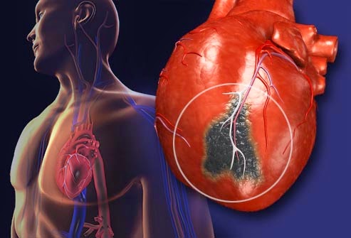
Usually the manifestations of heart failure begin insidiously, blurring at the outset with those of the underlying cardiac disorder and continuing gradually into a chronic state. However, the syndrome may also begin precipitously, as after myocardial infarction.
Compensatory ad|ustments are elicited by both acute and chronic heart failure. These include increase in peripheral vascular resistance, redistribution of blood flow, and increase in erythropoietic activity. But the adaptive mechanisms differ qualitatively, quantitatively, and often in direction. For example, acute distention of the left atnum generally promotes a sodium-poor diuresis, whereas chronic distention of the left atnum elicits salt and water retention. This distinction between acute and chronic heart failure is relevant to observations on experimental heart failure in animals, which is generally acute in onset and fulminating in course and usually ends abruptly with the death of the animal.
INITIATING MECHANISMS.
Each initiating mechanism generally imposes its own distinctive stamp upon the clinical syndrome. Thus the constellation of symptoms and signs originating in rheumatic heart disease differs from that caused by hypertensive heart disease; in turn, both have a different natural history from cor pulmonale. Indeed, even a single etiology, arteriosclerosis, may have distinctly different consequences, depending on the size and location of the vessels that are predominantly affected: progressive narrowing and gradual occlusion of peripheral branches of the coronary artenes may be so covert that the characteristic shortness of breath and undue fatigue of left ventricular failure may be misinterpreted as evidence of the general physical decline of advancing age. Conversely, abrupt closure of a ma)or coronary artery, as a consequence of thrombosis superimposed on an arteriosclerotic plaque, may elicit massive myocardial necrosis, followed by extensive myocardial scarring and unremitting heart failu
LEFT VS. RIGHT HEART FAILURE.
Although the ventricles have circumferential muscle and the ventricular septum in common, with rare exception, one ventricle fails before the other. Because of the prevalence of cardiac disorders which overload or damage the left ventricle, e.g., coronary arteriosclerosis and hypertension, heart failure usually begins with the left ventricle. Breathlessness, the key clinical expression of pulmonary congestion and edema, is the most common initial complaint. Conversely, when the right ventricle fails, systemic venous congestion and peripheral edema generally predominate. Left ventricular failure is the most common cause of night ventricular failure, and breathlessness often improves as right ventricular output falls and pulmonary congestion diminishes.
The mechanism by which left ventricular failure causes the right ventricle to fail is not clear. The conventional explanation puts the blame on pulmonary hypertension secondary to left ventricular failure. But the degree of pulmonary hypertension in this circumstance is rarely enough to constitute a formidable burden on the right ventricle. Much more likely as a predominant mechanism is interdependence of the two ventricles, which causes failure of the shared muscle, particularly in the ventricular septum. How often right ventricular failure causes left ventricular failure is still a matter of debate. Resolution of this problem is difficult because of the high frequency of independent and unrelated left ventricular disease in elderly patients with nght ventricular failure.
The combination of left and right ventricular failure, in which evidences of systemic and pulmonary venous hypertension predominate, is traditionally referred to as “congestive heart failure.” This is a colloquialism that conveys the image of severe physical incapacity, breathlessness, distended neck veins, hepatic engorgement, and troublesome peripheral edema. But it also highlights clinical features that can be measured at the bedside: a high venous pressure, a prolonged circulation time, and a diminished vital capacity, all of which improve as the patient gets better.
BACKWARD VS. FORWARD HEART FAILURE.
Somewhat reminiscent of the fable about the blind men and the elephant is the vintage controversy about “backward” versus “forward heart failure. “Backward failure” calls attention to the damming up of blood in the veins proximal to the failing ventricle and attributes to this venous congestion a critical role in the evolution of the syndrome of heart failure; “forward failure” assigns the same pivotal role to a decrease in cardiac output and hypoperfusion of organs. This distinction has little physiologic validity, because it is inevitable in a closed circuit that the inability of the heart to sustain its output (forward failure) and the pooling of blood on the venous inflow side (backward failure) must go hand in hand. However, this jargon often serves a useful practical purpose in highlighting clinical features.
LOW VS. HIGH OUTPUT FAILURE. Cardiac catheterization in man has made it possible to sort myocardial failure according to the level of the cardiac output. This practice has served at least three purposes: (1) to separate, on clinical and physiologic grounds, a type of myocardial failure (“high output failure”) in which the circulation remains vigorous and the extremities remain warm despite venous congestion, edema, and a lower cardiac output than existed prior to the heart failure; (2) to emphasize that the level of the cardiac output and the circulatory adjustments during myocardial failure are, to a large extent, a consequence of the cardiac output that existed prior to heart failure; and (3) to relate etiology and clinical evidences of heart failure to the state of the circulation: the more common causes of heart failure—arteriosclerosis, hypertension, myocardial disease, valvular disease, and pericardial disease—tend to be low output states; less common causes, such as hyperthyroidism,
Paget’s disease, anemia, beriberi, and arteriovenous fistula, tend to be high output states. But the hemodynamic hallmark of cardiac failure remains the same regardless of the level of cardiac output at rest: an inability of the heart to increase its output (or stroke volume or stroke work) as end-diastolic volume (as well as pressure) is increased.
The separation into “high” and “low” output failure is concerned with the clinical manifestations, rather than the causes, of myocardial failure. But it is also a hemodynamic reference to which the anatomist and the biochemist can relate the myocardial origins of heart failure. For example, “high output” failure probably has different biochemical bases from “low output” failure. Nor are all types of “low output” or “high output” failure apt to have the same biochemical origins. It is exceedingly unlikely that the “high output” failure of a peripheral arteriovenous fistula has the same anatomic or biochemical beginnings and evolution as the “high output” failure of severe anemia or malnutrition.
CONGESTIVE FAILURE VS. CONGESTED STATE.
Overfilling of the circulation, without myocardial failure, is the hallmark of the “congested state.” It is now common in intensive care facilities where massive infusions often represent last ditch efforts to combat systemic hypotension. Acutely, it is most often induced by rapid infusions; chronically, it is frequently encountered in severe anemia and in chronic renal insufficiency and less often in Paget’s disease or beriberi. In each of these situations, venous hypertension is a consequence of an expanded venous volume and heightened venomotor tone, rather than of heart failure.