Pinta disease is a chronic skin infection caused by Treponema carateum, giving rise to an initial lesion and a generalized secondary rash, both containing treponemes. Late skin manifestations comprise extensive dyschromic (treponeme-containing) and achromic (treponeme-free) conspicuous splotches. Antibodies are produced, detectable by serologic tests reactive also in syphilis and yaws. Organ systems are not involved, physical health is not impaired, and prenatal disease is not known.
History of Pinta Disease
Manifestations of pinta were described by Berochea and by Corona (1811). Frequent seroreactivity (Wassermann complement fixation test) in pinta patients led Menck (1926) to imply association with syphilis and made Herrejon11927) assume a treponeme to be the causative micro-organism. Armenteros and Triana (1938) identified T. carfiteum in Cuban pinta. Leon Blanco (1939) obtained early generalized eruptions by self-inoculation of. dark-field-controlled material from lesions. In therapy Corona (1811) showed the usefulness of mercury, Maria Graz (1913) of arsenobenzoles, and Varela (1944) of penicillin.
Etiology of Pinta Disease.
The author wishes to acknowledge the valuable advice, criticisms, and suggestions by his colleague Dr. S. Christiansen, Scientific Adviser, WHO Reference Centre for Treponematoses, State Serum Institute, Copenhagen, Denmark, concerning this article.
Epidemiology of Pinta Disease.
Pinta is an endemic treponematosis of large rural populations in tropical forest and valley regions of Central and South America. For example, in rural areas of Mexico the prevalence of pinta varied from 1.3 to 9 per cent (1964) of the census population. Pinta cases occasionally reported from Pacific islands, India, Indonesia, and West Africa have not been verified and may have been “pintide” yaws. Unlike yaws, pinta is not a disease of earliest childhood. Community data (Mexico) indicate age-specific prevalences of early pinta lesions to be 2.5 per cent below 5 years of age, 8.8 per cent between five and ten years, and 12.2 per cent between ten and fifteen years. In later life some early lesions occur also after 40 years of age.
Transmission presumably takes place by person-to-person contact and is facilitated by poor hygiene, low economic standards, and limited health services. Treponemes are present in early lesions as well as in extensive late dyschromic lesions, where they can be found up to 40 years after the infection. Pinta does not appear to be very contagious, because an infected spouse with treponema-containing lesions may sometimes not infect a serologically nonreactive partner or other family members. Scratches and insect bites may provide portals of entry for the agent. Blanco found 63 per cent of 257 initial lesions located on the legs and dorsum of feet. Arthropods (Simuliidae, Hip-pelates, or Ornithodorus) have not been convincingly demonstrated to be reservoirs of treponemes or biologic vectors, although mechanical transmission cannot be excluded.
Tie histopathology of pinta is characterized by a perivascular infiltrate of inflammatory cells composed of lymphocytes, some plasma cells, histiocytes. and macrophages. Swollen endothelial cells show no proliferation, arterioles and capillaries are not obliterated (as is the case in syphilis), and granulomatous tuberculoid structure is not observed. In dyschromic lesions accumulation of melanin-filled chromatophores in the upper corium :s characteristic, and is caused by pigment “fallout” from- the epidermis.
This might be a consequence of the primary process. In achromic lesions the picture is quite different—epidermis is atrophic with flattened rete pegs, melanocytes and melanin are lacking, elastic fibers are destroyed, and there is collagenic sclerosis, which explains the porcelain whiteness of achromic lesions. The pigment changes may result from direct action of T. carateum on the epidermal “melanin unit” (a melanocyte with a pool of associated malpighian cells [Duchon et al., 19681). Pinta lesions are localized to skin areas exposed to sunlight. This is unrelated to the number and distribution of melanocytes. A photosensitization process is therefore unlikely.
Clinical Manifestations and Diagnosis of Pinta Disease.
The incubation period in experimental pinta is 7 to 21 days. In man, the initial manifestation is a small papule developing by extension or by coalescence with satellite lesions into a scaly maculopapular lesion. There is regional lymphadenopathy. A generalized erythrosquamous rash develops three to nine months after infection, and can be of the “wandering” type. Palpable.polyadenitis is not a feature of secondary pinta in contrast to secondary syphilis. One to three years after the initial lesion, sizable dyschromic macules develop. These late lesions develop from secondary peptides or independently, and pass from slate blue through violet to brown and white — the final achromic phase of the pathogenic- process.
The time to pass these stages varies for different patches in the same individual, and the coexisting colored and white skin areas present a mottled appearance. Dyschromic lesions are usually located on frontal skin, cheeks, ears, forearms, back of hands, and dorsum of feet, but never the scalp. Blue lesions may be punctate, but most often appear as smudges on the brow, cheeks, and side of nose, and may last for one to two years. Brown lesions last much longer, and white elements are of lifelong duration. The achromic lesions are porcelain white, exhibit a “geographic coast” appearance; the skin is not supple and has no skin lines or lanugo. A different clinical course of pinta has been described in Cuba Pardo-Castello, 1942), where the early phase is limited to palms and soles, with hyperpigmented spots turning into keratotic elements.
The hyperpigmentation extends to the backs of hands and forearms. In Cuba implication of the cardiovascular and nervous systems has also been suggested. Such systemic involvement has not been verified in other pinta-affected areas of Central and South* America. For instance Mazotti (1966) found non-reactive treponemal antibody tests (TPI) in thecerebrospinal fluid of a series of advanced pinta patients in Mexico.
Early pinta may be difficult to differentiate from neurodermatitis (dark-field examination is decisive even if there is seroreactivity); trichophytosis of the glabrous skin (pinta does not have vesicles or pustules); pityriasis alba (early pinta is more infiltrated); tinea versicolor (secondary pintides are more sharply delineated and infiltrated); or psoriasis (on removal of scales in pinta no bleeding points appear on a smooth surface). In late pinta the leukoderma may closely resemble similar lesions in syphilis and yaws (scarring); vitiligo (supple skin in scalp and perianal areas); chloasma (pregnancy and disorders of female genitals); melanosis (teleangiectasis and poikiloderma are not features of pinta); or incontinentia pigmenti (urticaria prior to spots in infancy, pattern of splotches different, concomitant retinal and organ disease).
Laboratory Methods of Pinta Disease.
A diagnosis of pinta depends on microscopic dark-field demonstration of T. carateum in fluid from early initial and secondary generalized lesions and late dyschromic lesions, as well as on serological reagin and treponemal antibody tests. Methodologic considerations and interpretations concerning these are related to those in syphilis.
Treatment of Pinta Disease.
The treatment of choice is repository, long-acting penicillin, notably procaine penicillin G in oil with aluminium monostearate (PAM) and benzathine penicillin G (DBED). The considerations regarding time-dose relationship, oral therapy, and alternative antimicrobial drugs in persons hypersensitive to penicillin are the same as are discussed in the article on Syphilis. Rein et al. (1952) obtained highly satisfactory treatment results with 2.4 to 4.8 megaunits of PAM in early and late pinta. Injections of 2.4 megaunits of the longer-acting benzathine penicillin G can be effectively applied in a single dose or two injections of 1.2 megaunits in each buttock in one session.
Prognosis of Pinta Disease.
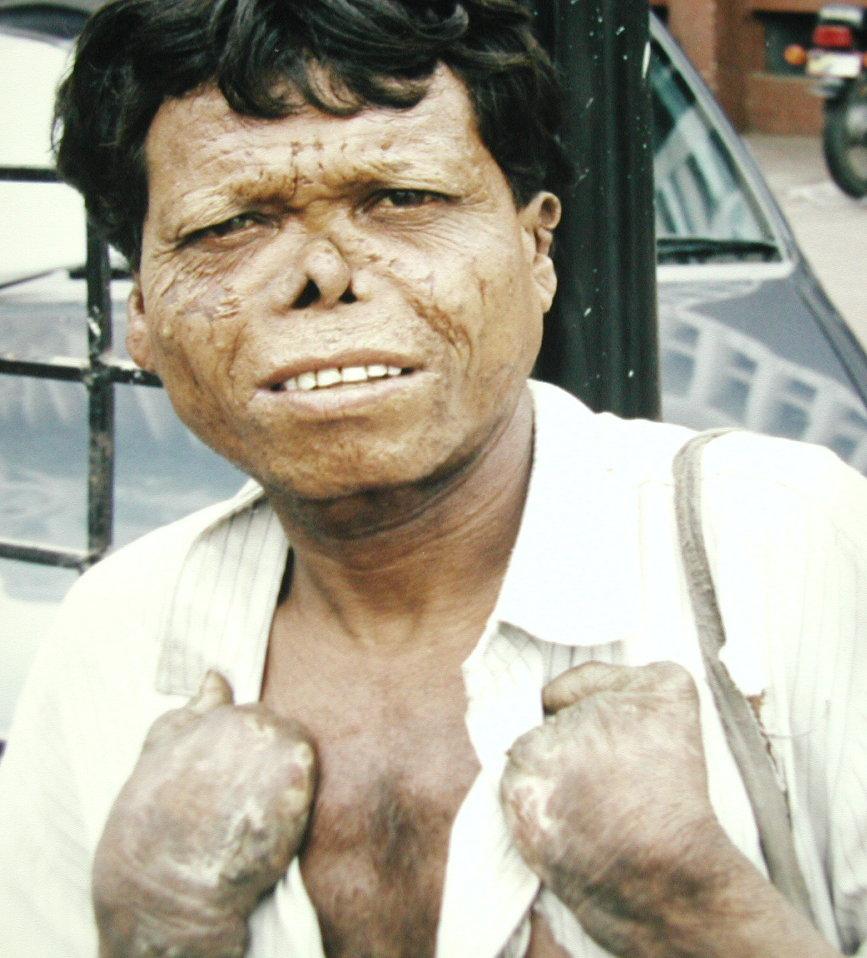
Pinta neither endangers life nor gives rise to prenatal disease. It has no appreciable effects on the general health of patients. Treatment causes treponemes to disappear rapidly from the lesions, which sometimes regress very slowly, depending on their extent. Achromic patches in which atrophy of the epidermis has occurred do not change. However, the disfigurement from pinta, notably of younger persons, is sometimes associated with psychologic misery and social ostracism. Freedom of choice of habitat, mate, and employment is curtailed.
Prevention of Pinta Disease.
Pinta prevention consists in examination and treatment of all cases and their contacts with long-acting penicillin, improvement of rural health services, hygiene, and economic standards. In the frame of the international treponematoses program of the World Health Organization (WHO) and the Pan-American Health Organization (PAHO) extensive penicillin treatment campaigns have been undertaken in rural endemic pinta areas by several national health administrations in Central and South America during the last two decades. For instance, in Mexico an estimated 350,000 pinta patients and contacts have been treated since 1959. Prevalence was reduced from 5.9 to 0.4 per cent in five main states.
Pinta disease works by causing a chronic bacterial infection in the body, primarily affecting the skin. Treponema carateum, the causative bacterium, enters the body through skin abrasions and triggers an immune response. This response, however, is unable to fully eliminate the bacteria. The result is the development of characteristic skin lesions and potential damage to other organs, such as the eyes. Early diagnosis and treatment are crucial to prevent the long-term effects of Pinta disease.