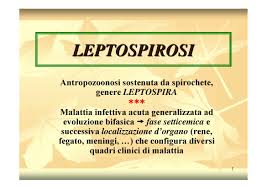
Leptospirosis is a broad term applied to all infections due to leptospires regardless of specific serotype. Correlation of clinical syndromes with infection by specific serotypes leads to the conclusion that a single serotype of Leptospira may be responsible for a variety of clinical features; likewise, a single syndrome, e.g., aseptic meningitis, may be caused by multiple serotypes of leptospires. Hence, there is preference for the general term leptospirosis rather than the multiple synonyms such as Weil’s disease, Canicola fever, etc.
Classification and Epidemiology.
The genus Leptospira contains only one species, L. interrogans, which may be subdivided into two complexes, interrogans and biflexa. The interrogans complex includes most of the pathogenic strains, whereas the biflexa complex includes mainly saprophytic strains with no recognized hosts. Within each complex, strains are classified by agglutination reactions into serogroups and serotypes. Despite contrary common usage, an example of the correct designation of leptospira is as follows: pomona serogroup of L. interrogans, not L. pomona. The interrogans complex now contains about 130 serotypes arranged in 16 serogroups (the number in parentheses refers to number of serotypes within the serogroup): icterohemorrhagiae (13), heb-domadis (28), autumnalis (13), canicola (11), australis (10), tarassovi or hyos (10), pyrogenes (9). bataviae (8), javanica (6), pomona (6), ballum (3), cynopteri (3), celledoni (2), gri’ppotyphosa (2), panama (2), and shermani (1). At least 22 serotypes of Leptospira occur naturally in the United States.
Infection in man is an incidental occurrence and is not essential to the maintenance of leptospiro-‘ sis in nature.. A wide range of domestic and wild animal hosts have been shown to have leptospirosis. In many species, such as opossums, skunks, raccoons, and foxes, infectivity ratios in the range or 10 to 50 per cent are not unusual. Interspecies spread of specific serotypes of leptospires between animal hosts is frequent, e.g., pomona, a serotype principally associated with livestock, has been demonstrated in dogs. The infection in animals may vary from inapparent illness to severe fatal disease. The carrier state may develop in many animals wherein the host may shed leptospires in its urine for months to years.
It has not been established that pathogenic leptospires are capable of multiplication outside a host. Survival in nature is governed by factors including pH of the urine of the host, pH of soil or water into which they are shed, and ambient temperature. Acid urine permits only limited survival; however, if the urine is neutral or alkaline and is shed into a similar moist environment which has low salinity and is not badly polluted with micro-organisms or detergents, and with a temperature above 22° C.^ leptospires may survive for several weeks. Human infections can occur either by direct contact with urine or tissue of an infected animal or indirectly through contaminated water or soil. The usual portals of entry in man are abraded skin, particularly about the feet, and exposed mucous membranes, conjunctival, nasal, and oral. The previously held concept that organisms could penetrate intact skin has been questioned. Although leptospires have been isolated from ticks, they appear to be unimportant in transmission.
With the ubiquitous infection of animals, leptospirosis in man can occur in all age groups, at all seasons, and in both sexes. However, it is primarily a disease of teen-age children and young to middle-aged adults (75 per cent of patients are between ages 10 and 50 years), occurs predominantly in males (90 per cent), and develops most frequently in hot weather (in the United States two thirds of infections occur from June to September). ThQ wide spectrum of animal hosts results in both urban and rural human disease. Leptospirosis has been considered an occupational disease; although up to one third of patients have direct contact with animals, e.g.’, farmers, abattoir workers, and veterinarians, the majority of patients have incidental exposure. Swimming or partial immersion in contaminated water has been implicated in one fifth of patients.
Pathology of Leptospirosis
In patients who have died with either hepatic involvement (Weil’s syndrome), renal involvement, or both, the significant gross changes include hemorrhages and bile-staining of tissues. The hemorrhages, which vary from petechial to ecchymotic, are widespread but are most prominent in skeletal muscle, kidneys, adrenals, liver, stomach, spleen, and lungs.
In skeletal muscle, focal necrotic and necrobiotic changes thought to be rather typical of leptospirosis occur. Biopsies early in the illness demonstrate swelling, vacuolation, and subsequently hyalinization. Leptospiral antigen has been demonstrated in these lesions by the fluorescent antibody technique. Healing ensues by the formation of new myofibrils with minimal fibrosis.
The changes, in the brain and meninges are also mini-tr.il and are not diagnostic. Thickening of the meninges with a polymorphonuclear leukocytic infiltration has been observed. Rarely, parenchymal lesions consisting of perivascular round-cell ;nitration may be encountered. Microscopic evidence of myocarditis, including focal hemorrhages, interstitial edema, and focal infiltration with lymphocytes and plasma cells, has been recorded. Pulmonary findings consist of a patchy, localized hemorrhagic pneumonitis. Special staining techniques utilizing silver impregnation methods have demonstrated organisms in the lumina of renal tubules but rarely in other organs.
Clinical Manifestations of Leptospirosis
General Features. The incubation period for icterohaemorrhagiae following immersion or accidental laboratory exposure has shown extremes of 2 to 20 days, the usual range being 7 to 13 days and the average, 10 days. This incubation, period does not vary significantly with other serotypes.
Leptospirosis is typically a biphasic illness.
The leptospiremic or first phase is characterized by manifestations of an acute infectious process. During this phase, leptospires are present in the blood and cerebrospinal fluid. The onset is typically abrupt (75 to 100 per cent of patients). Initial symptoms include headache, which- is usually frontal, less often retro-orbital, but occasionally may be bitemporal or occipital. Severe muscle aching occurs in most patients, the muscles of the thighs and lumbar areas being most prominently involved. The myalgia may be accompanied by extreme cutaneous hyperesthesia. Chills followed by rapidly rising temperature are prominent.
Following the abrupt onset, the leptospiremia phase typically lasts four to nine days. Features during this interval include recurrent chills, high spiking temperatures (usually 102° F. or greater), headache, and continued severe myalgia. Anorexia, nausea, and vomiting are encountered in one half or more of the patients. Pulmonary manifestations, usually either cough or chest pain, have varied in frequency of occurrence from less than 25 to 86 per cent. Hemoptysis occurs but is rare. Examination during this phase reveals an acutely ill febrile patient. A relative bradycardia may be noted. The blood pressure is usually normal, although European authors comment on early hypotension. Disturbances in sensorium may be encountered in up to 25 per cent of patients.