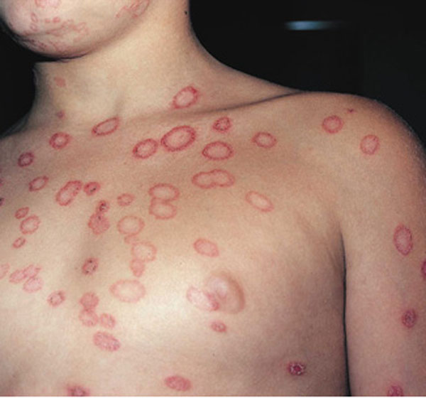
Infection with the guinea worm Disease usually presents as a skin ulceration at the site of emergence of the female adult worm. Man is probably the only reservoir, although monkeys and dogs can be experimentally infected. Humans infections are widespread in the tropics, occurring in local distribution in West Africa and :he Nile Valley, the Middle East, India and Pakistan, the Caribbean Islands, Guiana, and Brazil. Infection occurs on ingesting infected wateriness (Cyclops) present in drinking water from shallow wells or ponds.
The infective larvae in the Cyclops penetrate the intestinal walls and mature in the loose connective tissue under the skin, especially that of legs and feet. The male worm is small and dies after copulation. The female requires a year to become gravid, and then measures up to a meter long and is 2 mm. in diameter. When ready to discharge larvae, she approaches the skin surface, and a blister is produced by secretion of a :toxic substance from the anterior end of the worm. The blister breaks down to form an ulcer a few centimeters across, and the anterior end of the worm protrudes into this ulcer. On contact with water the head of the worm ruptures, and the uterus periodically discharges the tightly coiled larvae infective to the water flea. Secondary infection of the ulcer with resultant cellulites is common.
Guinea Worm Disease Symptoms In Humans.
Generalized allergic symptoms may occur prior to the blister formation or when surgical removal of the worm is attempted. Multiple infections are common. The lesion is usually on the .ower leg, but may occur on the genitalia, buttocks, or upper limbs. . In water carriers lesions have been observed on the back, suggesting that the worm is positively hydrotropic. Alternatively, the mature female may never reach the surface of the oody and may be absorbed or calcify in the tissue. The radiologic appearance is pathognomonic because the worm is so large. If a gravid worm dies in situ or is broken during extraction, cellulitis and secondary infection often occur. This may give rise to contractures. Also Clostridium tetani may contaminate the wound and tetanus may result. Rarely the adult worm involves serous cavities, the extradural space, or joints.
Guinea worm arthritis appears to be due to the presence of the adult worm or larvae in the joint. A microscopic diagnosis can be made by finding embryos in the exudate from the guinea worm ulcer after exposure to a few drops of water.Gradually winding the worm out of the ulcer by turning it on a stick a few centimeters a day is still common practice. Surgical extraction is also practiced. Recently niridazole (Ambilhar) has been reported to be lethal to the adult worm, which can readily be withdrawn after a course of 25 mg. per kilogram of body weight for seven days. Thiabendazole is also effective. Prevention involves constructing water sources that cannot be contaminated and killing cyclops by chlorination or boiling water to be used for drinking.
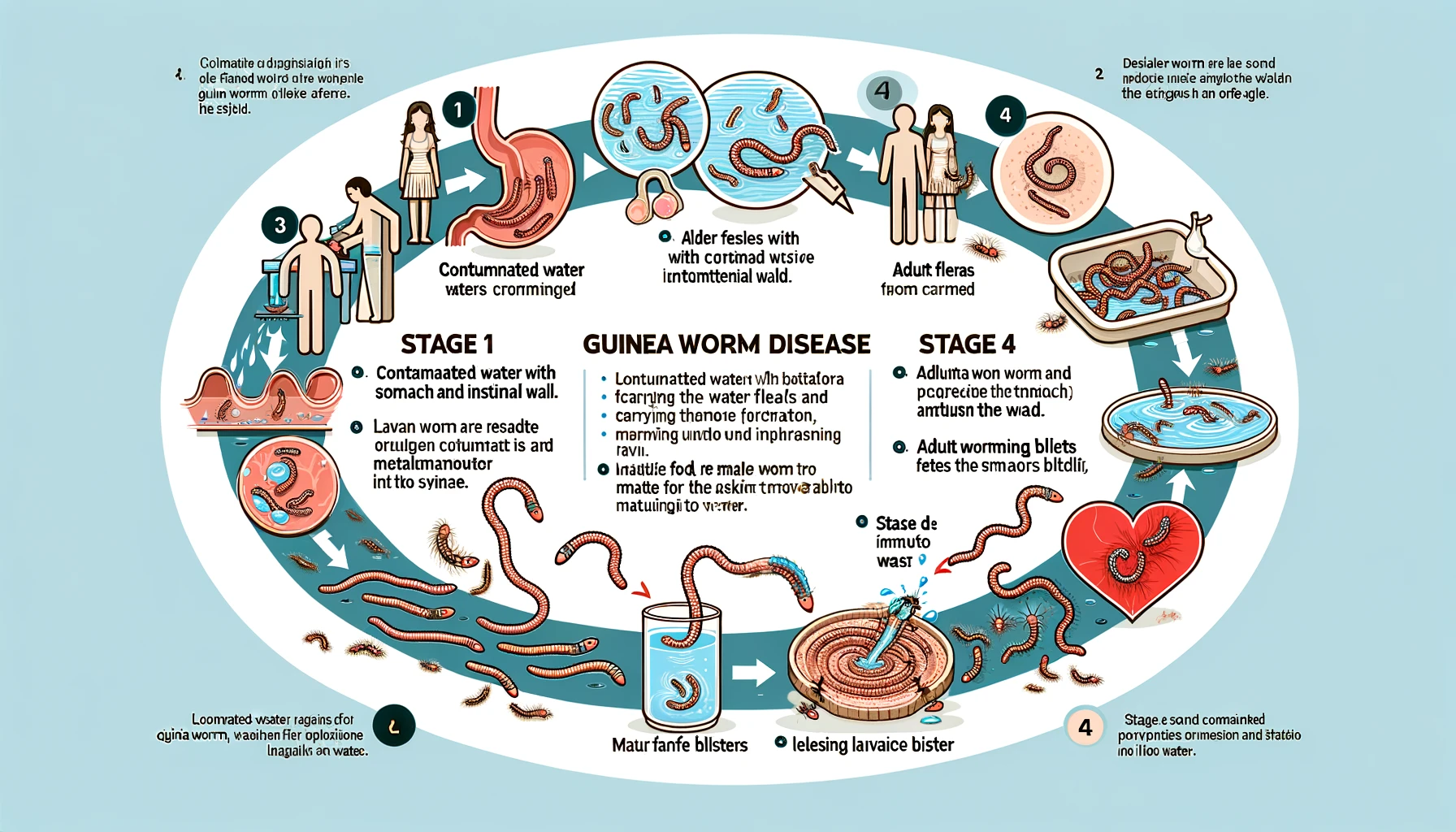
Guinea Worm Disease, also known as dracunculiasis, is a parasitic infection caused by the Guinea worm (Dracunculus medinensis). Here’s a guide in a tabular format explaining how this disease works:
| Stage | Description |
|---|---|
| 1. Contaminated Water Ingestion | The cycle begins when individuals drink water contaminated with copepods (tiny water fleas) that carry the Guinea worm larvae. |
| 2. Larvae Release and Maturation | Inside the human stomach, the copepods die, releasing the larvae, which then penetrate the stomach and intestinal wall, entering the abdominal cavity and retroperitoneal space. |
| 3. Reproduction and Migration | After about three months, the male and female worms mate. The male dies, and the female worm migrates, usually towards the lower limbs. |
| 4. Blister and Emergence | Approximately one year after infection, a painful blister forms on the skin, usually on the lower limb. This blister ruptures, exposing the worm. |
| 5. Worm Emergence and Water Contact | When an infected person immerses the blister in water (often due to pain relief), the worm begins to emerge from the wound and releases larvae back into the water, contaminating it. |
| 6. Ingestion by Copepods | The larvae are ingested by copepods, and after about two weeks inside the copepods, they become infectious to humans, thus completing the cycle. |
Prevention strategies include providing clean drinking water, filtering water through a fine cloth to remove copepods, and educating communities about avoiding water sources contaminated by the worm. There’s no vaccine or medication to treat Guinea Worm Disease; the worm must be gradually pulled out from the wound over several days to weeks.
Conclusion
Guinea worm disease is a painful and debilitating parasitic infection that affects millions of people in impoverished communities. Understanding how this disease works is crucial in developing effective prevention strategies and providing appropriate treatment. By improving access to clean water sources, promoting hygiene practices, and raising awareness, we can strive towards eradicating Guinea worm disease once and for all.