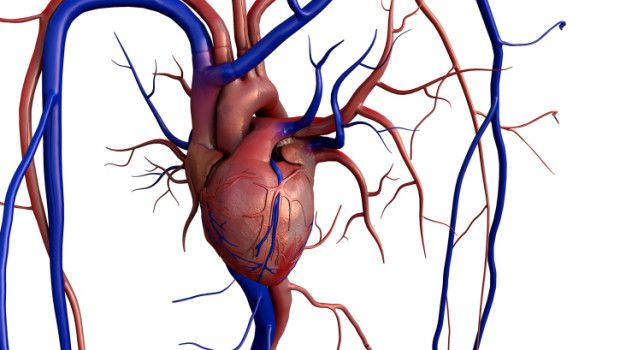
Eisenmenger syndrome is a complication of a birth defect that arises (congenital). A heart defect that causes a hole (shunt) to develop between two chambers of the heart. This hole causes blood to circulate abnormally in your heart and lungs.This syndrome is associated with marked elevation of pulmonar vascular resistance with reversed or bidirectional shunt, which is intracardiac or between the aorta and pulmonary arteries. Thus, pulmonary vascular disease is the hallmark of this syndrome and the site of the shunt is incidental. Medial hypertrophy of pulmonary arteries and arterioles is present and is associated with cellular, fibrotic, and fibroelastic intimal reactions, and in more severe forms plexi-form lesions encroach into the lumen of the vessel.

Symptoms OF Eisenmenger Syndrome
Eisenmenger syndrome signs and symptoms include:
- Bluish or grayish skin color (cyanosis)
- Large, rounded nails or feet (club)
- Easily tiring and lack of breath during the activity
- Shortness of breath at rest
- Chest pain or a sense of oppression
- Jump or run heart beats (palpitations)
- Fainting (syncope)
- Dizziness
- Numbness or tingling in fingers or toes
- Headach
The heart size is increased to a variable extent, greatest when there are shunts at the atrial level and when there is complicating tricuspid and’or pulmonary valve incompetence. The precordium is active with a right ventricular heave along the left sternal edge. Pulmonary arterial pulsations and the second heart sound may be palpable at the upper left sternal edge. The systolic murmur varies in intensity and is frequently initiated by a pulmonic ejection click.
The upper left sternal edge.
The systolic murmur varies in intensity and is frequently initiated by a pulmonic ejection click. The second heart sound is booming, single, or narrowly split in ventricular shunts, but wide, fixed splitting may be audible in isolated atrial shunts. Signs of pulmonary and/or tricuspid valve regurgitation are superimposed when there is dilatation of these valve rings secondary to pulmonary hypertension or right ventricular failure. The electrocardiogram shows marked right ventricular or biventricular hypertrophy with prominent P waves. Complete right bundle branch block may be present, especially when the shunt is at the atnal level. In others the electrocardiogram is influenced by the underlying anomaly (e.g., single ventricle, ventricular inversion, etc.). The chest roentgenogram confirms the degree of cardiomegaly.
The pulmonary trunk is enlarged with prominence of the primary divisions, which diminish in caliber in the penpheral branches. The echocardiogram helps to identify the anatomy of the underlying intracardiac or extracardiac malformation. Cardiac catheterization is undertaken when the diagnosis cannot be established by clinical findings and noninvasive studies. One of the purposes of catheterization is to determine whether the pulmonary vascular bed is vasoactive, as indicated by a fall in pulmonary artery pressure and resistance during the breathing of 100 percent oxygen. Another major indication is to exclude the presence of left ventncular inflow lesions, which result in elevation of pulmonary venous pressure and secondary pulmonary hypertension. Angiocardiography carries a small increased risk because the contrast medium may produce a fall in systemic.
Polycythemia may become extreme, and when the hematocrit exceeds 70 per cent excruciating headaches may occur. These are difficult to treat but do respond to repeated venesection. This treatment should not be undertaken lightly, since reduction of red cell count and blood volume is not tolerated. Heart rate and blood pressure are monitored during the procedure. Small aliquots of blood (♦ 30 ml) are removed and immediately replaced with a similar volume of fresh frozen plasma or human albumin. Repeated venesection results in iron deficiency anemia so that daily oral iron replacement is essential. The usual goal is to reduce the hematocrit to between 55 and 60 per cent. Hemoptysis occurs from rupture of pulmonary vessels or is due to pulmonary arterial thrombosis or embolism.
This symptom is usually limited to adult life, blood loss is not excessive, and symptomatic treatment is all that is needed. However, hemoptysis can be life threatening if associated with hypotension, an increase in the degree of hypoxemia, and the development of acidemia. Long-term anticoagulation is not indicated. Syncope and sudden death cannot be predicted, but patients with Eisen-menger syndrome between the ages of about 20 and 40 years are at risk. The mechanism is not clear but has been attributed to arrhythmias, probably ventricular tachyarrhythmias, which result in hypotension and an increase in right to left shunting. Pregnancy is not well tolerated and sudden death has been reported during the third trimester or in the postpartum period.
How Eisenmenger’s Syndrome develops
For most people who have Eisenmenger’s syndrome, the cause of their condition is due to a hole (shunt) between the major blood vessels or chambers of your heart. This shunt is a cardiac defect we are born (congenital). Cardiac defects that can cause Eisenmenger’s syndrome include:
- Ventricular septal defect. This shunt in the wall of tissue that divides the left and right sides of the main pumping chambers of the heart (ventricles) is the most common cause of Eisenmenger’s syndrome.
- Defect of atrial septum. An atrial septal defect is a shunt in the tissue wall that divides the left and right sides of the upper chambers of the heart (atria).
- Patency of the arterial duct. This heart defect is an opening between the pulmonary artery that carries oxygen-poor blood to the lungs and the artery that carries oxygen-rich blood to the rest of your body (aorta).
- Defect of the atrioventricular canal. In the heart defect, there is a large hole in the center of the heart where the walls between the upper chambers (atriums) and the lower chambers (ventricles) meet. Furthermore, some of the valves in the heart can not function properly.
Eisenmenger Syndrome Treatment.
Surgical treatment of the cardiac anomaly is contraindicated because these patients succumb to the effects of pulmonary vascular disease. Palliation has been successful in the presence of transposition of the great artenes, ventricular septal deffect, and severe pulmonary vascular disease; the procedure involves redirection of the venous return (as described under transposition of the great arteries), but the ventricular defect is not closed. The experience with transplantation of the heart and lungs is still small and follow-up is short, but this therapy is being watched with interest, since patients with progressive symptoms are at great risk of dying. Drugs have been used to attempt to manipulate pulmonary and systemic vascular resistance to reduce the right to left shunt; generally the results have been disappointing.
Prevention;What can you do After Having Eisenmenger Syndrome.
- Take note of any previous cardiac treatments. Since Eisenmenger’s syndrome most often develops as a complication of a heart defect, it’s important that your doctor about any medications you’ve taken, or surgeries or procedures you’ve had if you’ve previously been diagnosed with a heart defect.
- Be aware of any pre-appointment restrictions. When making the appointment, be sure to ask if there is anything you need to do in advance, such as filling out forms or restricting your diet. For some imaging tests, for example, it may be necessary to fast for a period of time in advance.
- Write down all the symptoms you have encountered, including those that may seem unrelated to Eisenmenger’s syndrome. Try to remember when they started. Be specific, such as days, weeks, months, and try to avoid vague terms like “a little while ago.”
- Write down key personal information, including a family history of heart defects, pulmonary hypertension, lung disease, heart disease, stroke, high blood pressure or diabetes, and any major stresses or recent life changes.
- Make a list of all the medications, as well as vitamins or supplements you are taking. Also, be sure to tell your doctor if you have recently stopped taking medication.
- Take a family member or friend, long, if possible. It can sometimes be difficult to remember all the information provided during an appointment. Someone who accompanies you can remember something that you have lost or forgotten.