Echocardiogram is an ultrasound examination of the heart that provides images obtained through sound. Our body has different tissues and with it different degrees sound waves.When the ultrasound device emits high frequency sound (over 20,000 cycles per second), far beyond human hearing capacity, it captures the echo return and transforms into a black and white image for the physician to analyze.
Echocardiography comprises a group of diagnostic procedures in which ultrasound is used to examine the heart. Ultrasound is sound with a frequency above the audible range. The upper limit of audible frequencies is approximately 20,000 cycles per second. The frequency used for echocardiography ranges from 1 million to 7 million cycles per second or 1 to 7 mega-Hertz. The term echocardiography comes from the fact that a cardiac image is obtained by recording the reflected sound waves or echoes of a beam of ultrasound aimed at the heart.
A burst of ultrasound is produced by a transducer, and the sonic waves travel from the transducer much as a light beam from a flashlight. When the ultrasonic beam strikes an interface between two media that have different acoustic properties, such as a difference in density, the ultrasound is reflected and refracted just as when a light beam strikes a reflecting surface such as water. If the reflecting surface is perpendicular to the transducer, some of the reflected ultrasonic waves or echoes will retrace their path and strike the transducer.
If one knows the velocity at which sound travels in the medium being examined and if one knows that it takes for a burst of ultrasound to leave the transducer, the reflecting object, and return as an echo, one can calculate the distance of the reflecting surface from the transducer. Since the velocity of sound transmission in human soft tissue is known, echo-cardiographs are calibrated for the velocity of sound in this medium, and distances between reflecting structures and the transducer are automatically indicated on the oscilloscope.
Only part of the ultrasonic energy is reflected by any given acoustic interface. The ultrasound that continues through the initial reflecting surface will strike any deeper interfaces and again be reflected back to the transducer. Since the transit time is longer, the deeper interface will be displayed on the oscilloscope as being farther from the transducer. The ultrasound is transmitted in short bursts or ultrasonic pulses at a rate of 1000 per second or more. This rapid sampling rate permits an accurate recording of cardiac motion.
Although echocardiography is usually noninvasive, it is possible to obtain additional diagnostic information by a peripheral venous injection of almost any liquid Such injections create tiny suspended bubbles of air by a process of cavitation, and these bubbles produce a mass of echoes within the heart. These tiny bubbles do not pass through capillaries. The combination of such injections and echocardiography is known as contrast echocardiography. This technique is particularly helpful in detecting intracardiac shunts and tricuspid regurgitation. Such injections can also be made through catheters in the cardiac catheterization laboratory to obviate or supplement selective angiography. Of course, such a use of echocardiography is no longer noninvasive.
Ultrasound can also provide diagnostic information utilizing the Doppler principle. This principle states that if a sound beam strikes an interface that is moving in the axis of the beam, the motion of that reflecting surface will alter the frequency of the reflected sound The amount of frequency change is a function of the velocity with which the reflecting object is moving. When dealing with ultrasound the difference in frequency or Doppler shift is in the audible range. Thus, when an ultrasonic beam strikes a reflecting surface which moves, an audible Doppler signal is produced. Doppler ultrasound has been used most extensively by examining moving columns of blood. The reflecting interfaces are red blood cells.
What are the types of echocardiogram ?
- One-dimensional echocardiogram : first version of the echocardiogram , used mainly to measure the diameters of the cardiac chambers and the myocardial thickness.
- Two-dimensional echocardiogram : allows the transformation of the images into dimensional figures allowing a better anatomical evaluation.
- Transthoracic echocardiogram : is the traditional and most common type of echocardiogram in which the transducer, which emits and captures sound waves for imaging, is slid over the cardiac region ofthe patient’s chest.
- Echocardiography transesophageal : the ultrasound transducer, high frequency, is glued inside theesophagus , the height of the heart through an optical fiber probe. The technique allows a closer examination of certain specific cardiac structures, because of the greater proximity of the transducer to the heart .
- Pharmacological stress echocardiography : ultrasonography of the heart with venous infusion of cardiac “stress” drugs, which increase the body’s oxygen consumption and whose responses are adequately monitored.
- Echocardiography fetal : performed through the abdominal wall of the pregnant and directed to the heart of the fetus in pregnancy, allows to evaluate the heart intra uterus .
- Echocardiogram with Doppler : allows to evaluate the circulatory conditions inside the heart.
What Diseases Are Seen With The Echocardiogram?
Objectively we can list the following diseases:
- Heart failure, known as weak heart
- Diseases of the heart valves, known as blow
- Birth defects, known as congenital
- Diseases of the pericardium, covering the heart
- Diseases of the thoracic aorta, known as dilated aorta
- Cardiac tumors, benign as atrial myxoma
- Growth of atrial and ventricular cavities
- Presence of clots within the heart that can migrate to the body.
How is the echocardiogram performed?
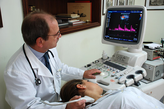
The standard examination is simple, painless and usually rapid, not requiring pre-treatment. The patient should lie down on a stretcher, on his or her side, with the echo cardiograph next to it. The doctor will place on your chest some electrodes. It is fixed to the skin by rubber “suction cups” and will slide through your chest a transducer that emits high frequency sound waves, and is refracted in the heart.On the exam, the doctor will try to direct the sound beam to the cardiac structures that you want to examine. This will form an image as seen below.
Because it is an examination that has no side effects , It is relatively inexpensive and easy to operate transport, and excellent diagnostic range , echocardiography is of great importance in modern cardiology.