Chromosomal abnormalities occur with greatly different frequencies at various times in life. The highest frequency and the greatest variety are found among Hponlaneous abortuses, 30 to 40 percent of whom have a major chromosomal defect. Among livehorn infants, about (i per 1000 have a chromosomal defect sufficiently severe tocauscdisabilit at some time in their lives. Alniut half of these abnormalities involve the autosomes. whereas the remainder are sex chromosomal disorders. Among adults in the general population, the abnormalities are largely con fined to the sex chromosomal disorders.
There are clinically definable subgroups within these population segments that have an increased risk for a chromosomal disorder Infants with multiple congenital defects, for example, have a 5 to 10 per cent probability of possessing a major autosomal abnormality. The occurrence in a family of individuals with a similar group of congenital malformations is often the result of an inheritable chromosomal disorder Such families may also have an increased number of spontaneous abortions Sex chromosomal disorders occur with increased frequency among infertile males and among women with primary amenorrhea They are also found to In- unusually frequent among males incarcerated in penal institutions.
Preparation of Cell for Chromosomal Analysis.
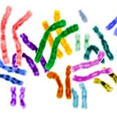
The chromosomes in the non-dividing inter-phase cell an so extended and intertwined that they cannot he individually distinguished. During cell division <miti>siN>, however. they become greatly condensed and are then visible as separate entities Dividing cells can be arrested in that phase of mitosis (metaphase) at which the chromosome* are most contracted by treatment with colchicine By the use of colchicine and by swelling the cells in hypotonic solution to increase the separation of the chromosomes. preparations can be produced in which each chromosome is clearly visible
Dividing cells can be obtained directly from bone marrow or by culturing a small piece of skin The procedure required for obtaining either of these specimens, however. is unpleasant for the patient. In addition, skin cells must be cultured for several weeks before a sufficient number of dividing cells is present. Fortunately, the normally nondividing leukocyte from peripheral blood can be stimulated to divide when exposed to phytohemagglutinin 1PHA1 PH A appears to act only on lymphocytes • specifically, thymus-dependent lymphocytes!. Stimulated lymphocytes undergo a transient wave of mitosis that reaches a peak in three to four days By a combination of 1*11 A. colchicine, and hypotonic treatments, suitable chromosome preparations can be obtained from a few drops of capillary blood.
In view of the simplicity of the procedure, most chromosomal analyst’s are now carried out with peripheral blood specimens. Hone marrow cells may nonetheless be used in the study of diseases, such as myelogenous leukemia, which are confined to these cells Skin cells may be examined when a difference between the skin cell and lymphocyte chromosome complement is suspected. or when blood samples cannot be obtained, as in abortuses.
Amniocentesis.
The amniotic fluid comprises a special source of fetal cells which has attained great practical importance in recent years. Ten to 20 ml of fluid can be withdrawn with apparent safety as early as the fourteenth to fifteenth week of gestation. The amniotic cells may Ik- examined directly for sex determination by staining for the Harr body (sex chmmatin mass) or for the Y body, as will be described later Mon- commonly, they are cultured and the dividing cells are treated to obtain metaphase preparations by the method previously mentioned. Amniotic cells are unusual in that tetraploid cells (cells with two complete chromosomal complements) occur with significant frequency even when the fetus is cytologically normal. This may indicate that amniotic cells may arise in part from the fetal memhranes and may not be totally representative of the fetus proper Chromosomal examination of the fetus may alter in the future if attempts to obtain fetal blood or skin biopsies through an amnioscope are successful.
Amniocentesis for chromosomal analysis can he considered for those pregnancies in which the risk of a chromosomal ly abnormal fetus is thought to be significantly greater than the risk of damage to the fetus from the amniocentesis procedure itself.
Nomenclature for Chromotomal Abnormalities.
In the shorthand notation now generally U8ed to describe the chromosome complement of an individual, the number of chromosomes is specified first, followed by the listing of the sex chromosomes In this notation the normal female karyotype is 46.XX and the normal male is 46,XY. Any deviations from the normal karyotype are written after the sex chromosome listing. An individual autosome is referred to by number, its upper (shorter! arm by p and its lower arm by q. A plus or a minus sign written after the p or q indicates an increase (+> or decrease (—) in length of the arm When written before a designated chromosome, the sign indicates that the chromosome is extra (+) or missing (— >. (Examples: 46.XY.18q describes a male with 46 chromosomes, including one chromosome 18 whose long arm is diminished in length. 47,XX.+21 describes a female with 47 chromosomes, including an extra chromosome 21 in addition to the 46 chromosome* of the normal kuryotype.
The Barr Body end the Y Body.
Despite the detaililed by tin- various banding techniques none is able to distinguish between the two X chromosomes of the normal female It is known that only one X chromosome is active in any cell, each X chromosome in excess of one condenses to form a sex chromatin mass iBarr body) visible at the periphery of the nucleus. The number of X chromosomes may be indirectly determined by examination of buccal mucosal cells for the number of Barr bodies.
Condensed, inactive X chromosomes an- also delayed in the timing of their DNA synthesis, relative to the active X chromosome The late-replicating X chromosome can he identified by autoradiography using Intuited thymidine.
In over 99 percent of males, the distal part of the long arm of the Y chromosome fluoresces brilliantly after staining with quinacrine. The intensity is sufficiently great that this region can be seen as a spot of fluorescence, the Y body, in the interphase nucleus Y-bearing cells can thereby be recognized in cells from blood, hair follicles, and tissue sections, and even in sperm.
Polymorphic Variations in the Human Karyotype.
In addition to those alterations in the karyotype which are directly or potentially associated with disease, some variations are known which are apparently without consequence to the individual who possesses them or to his progeny. These polymorphic vanations. as they are called, may represent changes in the DNA sequence of the chromosomal region concerned They are transmitted to progeny as part of the chromosome which contains them The more commonly n*cognized variations of this type occur in the length of the short arms of the acrocentric chromosomes, t he size of their satellites, the length of the fluorescent segment in the long arm of the Y. and the length of the secondary constrictions of 1, 9, and 16.

